8SJ4 | pdb_00008sj4

8F3-1H9-Ara h 6


Experimental Data Snapshot

  • Method: X-RAY DIFFRACTION
  • Resolution: 2.67 Å
  • R-Value Free: 
    0.232 (Depositor), 0.230 (DCC) 
  • R-Value Work: 
    0.190 (Depositor), 0.190 (DCC) 
  • R-Value Observed: 
    0.192 (Depositor) 

Starting Models: experimental
View more details

wwPDB Validation   3D Report Full Report


This is version 1.1 of the entry. See complete history


Literature

Structural determinants of peanut induced anaphylaxis.

Smith, S.A.Shrem, R.A.Lanca, B.B.C.Zhang, J.Wong, J.J.W.Croote, D.Peebles Jr., R.S.Spiller, B.W.

(2025) J Allergy Clin Immunol 

  • DOI: https://doi.org/10.1016/j.jaci.2024.12.1095
  • Primary Citation of Related Structures:  
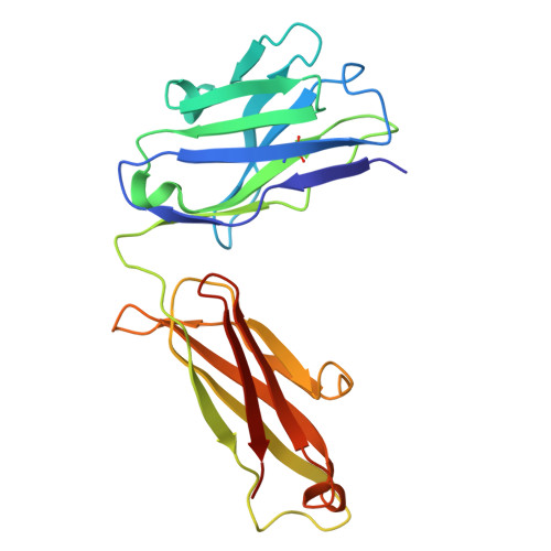
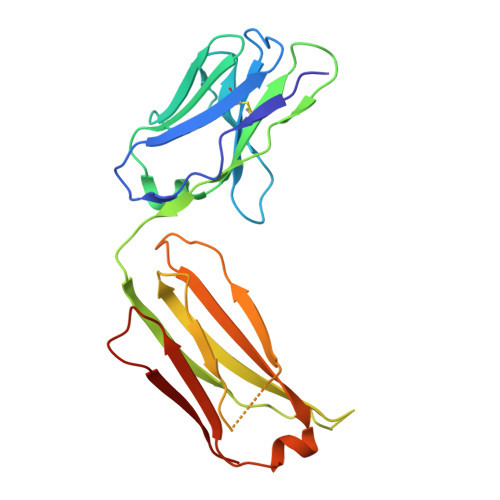
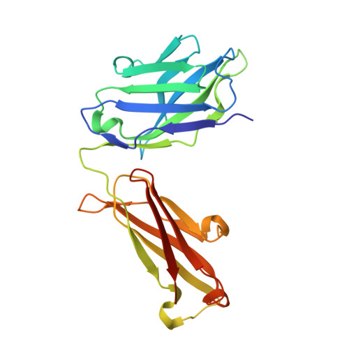
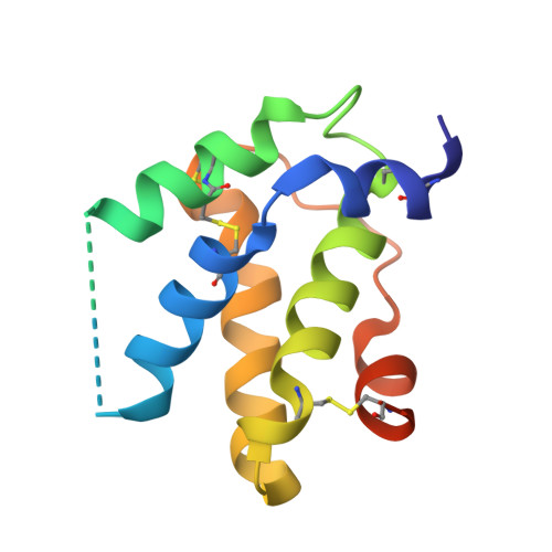
    8SHV, 8SHW, 8SI1, 8SJ4, 8SJ6, 8SJA

  • PubMed Abstract: 

    Human monoclonal IgE antibodies recognizing peanut allergens have recently become available, but we lack a detailed understanding of how these IgEs target allergens. To determine the molecular details of the antibody-allergen interaction for a panel of clinically important human IgE monoclonal antibodies and to develop strategies to disrupt disease causing antibody-allergen interactions. We identified candidates from a panel of epitope binned human IgE monoclonals that recognize two important and homologous peanut allergens, Ara h 2 and Ara h 6. Crystal structures were determined revealing the interfaces (antigenic sites) of exemplars of five common IgE bins. Among the common antigenic sites on Ara h 2 and Ara h 6, two sites (A and B) are highly conserved between the allergens, explaining the cross reactivity of antibodies that recognize these sites. Three sites (C, D, and F) involve residues that are not conserved between the allergens. Of the five common sites, three Sites (B, C, and D) involve residues that are only near each other when the allergens are properly folded, such that these sites are conformational. Two additional sites (sites A and F) involve largely linear stretches of amino acids. Site F targeted antibody, 38B7, binds to a peptide sequence DPYSP OH S, in which hydroxylation of the last proline is critical for binding. This sequence is repeated two or three times depending on the Ara h 2 isoform, enabling 38B7 to induce anaphylaxis as a single monoclonal, without a second antibody. We have mutated key residues in each site and created a panel of hypoallergens, having reduced IgE mAb binding and lacking the ability to induce anaphylaxis in our murine model. We created a structural map of the IgE antibody response to the most important peanut allergen proteins to enable the design of new allergy immunotherapies and vaccines.


  • Organizational Affiliation
    • Department of Medicine, Vanderbilt University Medical Center, Vanderbilt University, Nashville, TN; Department of Pathology, Microbiology and Immunology, Vanderbilt University Medical Center, Vanderbilt University, Nashville, TN.

Macromolecules
Find similar proteins by:  (by identity cutoff)  |  3D Structure
Entity ID: 1
MoleculeChains Sequence LengthOrganismDetailsImage
1H9 heavy chainA [auth H]218Homo sapiensMutation(s): 0 
Entity Groups  
Sequence Clusters30% Identity50% Identity70% Identity90% Identity95% Identity100% Identity
Sequence Annotations
Expand
  • Reference Sequence
Find similar proteins by:  (by identity cutoff)  |  3D Structure
Entity ID: 2
MoleculeChains Sequence LengthOrganismDetailsImage
1H9 light chain kappaB [auth L]214Homo sapiensMutation(s): 0 
Entity Groups  
Sequence Clusters30% Identity50% Identity70% Identity90% Identity95% Identity100% Identity
Sequence Annotations
Expand
  • Reference Sequence
Find similar proteins by:  (by identity cutoff)  |  3D Structure
Entity ID: 3
MoleculeChains Sequence LengthOrganismDetailsImage
8F3 light chain kappaC [auth D]214Homo sapiensMutation(s): 0 
Entity Groups  
Sequence Clusters30% Identity50% Identity70% Identity90% Identity95% Identity100% Identity
Sequence Annotations
Expand
  • Reference Sequence
Find similar proteins by:  (by identity cutoff)  |  3D Structure
Entity ID: 4
MoleculeChains Sequence LengthOrganismDetailsImage
8F3 heavy chainD [auth F]225Homo sapiensMutation(s): 0 
Entity Groups  
Sequence Clusters30% Identity50% Identity70% Identity90% Identity95% Identity100% Identity
Sequence Annotations
Expand
  • Reference Sequence
Find similar proteins by:  (by identity cutoff)  |  3D Structure
Entity ID: 5
MoleculeChains Sequence LengthOrganismDetailsImage
ConglutinE [auth A]122Arachis hypogaeaMutation(s): 0 
UniProt
Find proteins for Q647G9 (Arachis hypogaea)
Explore Q647G9 
Go to UniProtKB:  Q647G9
Entity Groups  
Sequence Clusters30% Identity50% Identity70% Identity90% Identity95% Identity100% Identity
UniProt GroupQ647G9
Sequence Annotations
Expand
  • Reference Sequence
Experimental Data & Validation

Experimental Data

  • Method: X-RAY DIFFRACTION
  • Resolution: 2.67 Å
  • R-Value Free:  0.232 (Depositor), 0.230 (DCC) 
  • R-Value Work:  0.190 (Depositor), 0.190 (DCC) 
  • R-Value Observed: 0.192 (Depositor) 
Space Group: P 1 21 1
Unit Cell:
Length ( Å )Angle ( ˚ )
a = 39.382α = 90
b = 85.45β = 96.17
c = 165.831γ = 90
Software Package:
Software NamePurpose
PHENIXrefinement
autoPROCdata processing
XDSdata reduction
Aimlessdata scaling
PHASERphasing

Structure Validation

View Full Validation Report


Entry History & Funding Information

Deposition Data


Funding OrganizationLocationGrant Number
National Institutes of Health/Eunice Kennedy Shriver National Institute of Child Health & Human Development (NIH/NICHD)United States5R01AI155668

Revision History  (Full details and data files)

  • Version 1.0: 2024-12-25
    Type: Initial release
  • Version 1.1: 2025-01-29
    Changes: Database references