8VCL | pdb_00008vcl

Crystal structure of HLA-A*03:01 in complex with a mutant PIK3CA peptide

  • Classification: IMMUNE SYSTEM
  • Organism(s): Homo sapiens
  • Expression System: Escherichia coli
  • Mutation(s): Yes 

  • Deposited: 2023-12-14 Released: 2024-12-11 
  • Deposition Author(s): Ma, J., Baker, B.M.
  • Funding Organization(s): National Institutes of Health/National Institute of General Medical Sciences (NIH/NIGMS)

Experimental Data Snapshot

  • Method: X-RAY DIFFRACTION
  • Resolution: 2.40 Å
  • R-Value Free: 
    0.230 (Depositor), 0.230 (DCC) 
  • R-Value Work: 
    0.194 (Depositor), 0.194 (DCC) 
  • R-Value Observed: 
    0.197 (Depositor) 

Starting Model: experimental
View more details

wwPDB Validation   3D Report Full Report


This is version 1.1 of the entry. See complete history


Literature

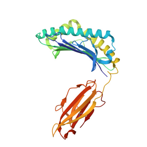
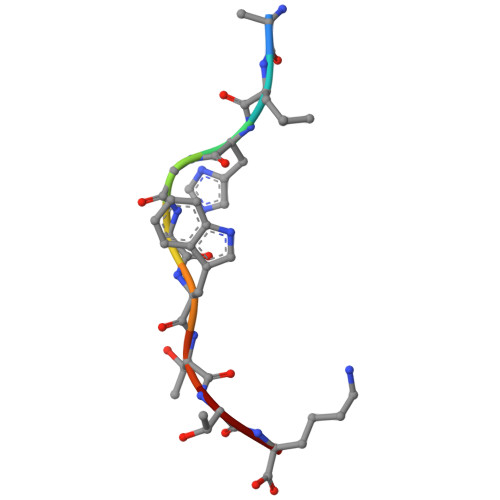
Dynamic allostery in the peptide/MHC complex enables TCR neoantigen selectivity.

Ma, J.Ayres, C.M.Brambley, C.A.Chandran, S.S.Rosales, T.J.Perera, W.W.J.G.Eldaly, B.Murray, W.T.Corcelli, S.A.Kovrigin, E.L.Klebanoff, C.A.Baker, B.M.

(2025) Nat Commun 16: 849-849

  • DOI: https://doi.org/10.1038/s41467-025-56004-8
  • Primary Citation of Related Structures:  
    8VCL, 9ASG

  • PubMed Abstract: 

    The inherent antigen cross-reactivity of the T cell receptor (TCR) is balanced by high specificity. Surprisingly, TCR specificity often manifests in ways not easily interpreted from static structures. Here we show that TCR discrimination between an HLA-A*03:01 (HLA-A3)-restricted public neoantigen and its wild-type (WT) counterpart emerges from distinct motions within the HLA-A3 peptide binding groove that vary with the identity of the peptide's first primary anchor. These motions create a dynamic gate that, in the presence of the WT peptide, impedes a large conformational change required for TCR binding. The neoantigen is insusceptible to this limiting dynamic, and, with the gate open, upon TCR binding the central tryptophan can transit underneath the peptide backbone to the opposing side of the HLA-A3 peptide binding groove. Our findings thus reveal a novel mechanism driving TCR specificity for a cancer neoantigen that is rooted in the dynamic and allosteric nature of peptide/MHC-I binding grooves, with implications for resolving long-standing and often confounding questions about T cell specificity.


  • Organizational Affiliation
    • Department of Chemistry and Biochemistry, University of Notre Dame, Notre Dame, IN, USA.

Macromolecules
Find similar proteins by:  (by identity cutoff)  |  3D Structure
Entity ID: 1
MoleculeChains Sequence LengthOrganismDetailsImage
HLA class I histocompatibility antigen, A alpha chain274Homo sapiensMutation(s): 0 
Gene Names: HLA-AHLAA
UniProt & NIH Common Fund Data Resources
Find proteins for P04439 (Homo sapiens)
Explore P04439 
Go to UniProtKB:  P04439
PHAROS:  P04439
GTEx:  ENSG00000206503 
Entity Groups  
Sequence Clusters30% Identity50% Identity70% Identity90% Identity95% Identity100% Identity
UniProt GroupP04439
Sequence Annotations
Expand
  • Reference Sequence
Find similar proteins by:  (by identity cutoff)  |  3D Structure
Entity ID: 2
MoleculeChains Sequence LengthOrganismDetailsImage
Beta-2-microglobulin100Homo sapiensMutation(s): 0 
Gene Names: B2MCDABP0092HDCMA22P
UniProt & NIH Common Fund Data Resources
Find proteins for P61769 (Homo sapiens)
Explore P61769 
Go to UniProtKB:  P61769
PHAROS:  P61769
GTEx:  ENSG00000166710 
Entity Groups  
Sequence Clusters30% Identity50% Identity70% Identity90% Identity95% Identity100% Identity
UniProt GroupP61769
Sequence Annotations
Expand
  • Reference Sequence

Find similar proteins by:  Sequence   |   3D Structure  

Entity ID: 3
MoleculeChains Sequence LengthOrganismDetailsImage
Mutant PIK3CA peptide9Homo sapiensMutation(s): 1 
EC: 2.7.1.137 (UniProt), 2.7.11.1 (UniProt), 2.7.1.153 (UniProt)
UniProt & NIH Common Fund Data Resources
Find proteins for P42336 (Homo sapiens)
Explore P42336 
Go to UniProtKB:  P42336
PHAROS:  P42336
GTEx:  ENSG00000121879 
Entity Groups  
UniProt GroupP42336
Sequence Annotations
Expand
  • Reference Sequence
Experimental Data & Validation

Experimental Data

  • Method: X-RAY DIFFRACTION
  • Resolution: 2.40 Å
  • R-Value Free:  0.230 (Depositor), 0.230 (DCC) 
  • R-Value Work:  0.194 (Depositor), 0.194 (DCC) 
  • R-Value Observed: 0.197 (Depositor) 
Space Group: P 6 2 2
Unit Cell:
Length ( Å )Angle ( ˚ )
a = 156.42α = 90
b = 156.42β = 90
c = 85.863γ = 120
Software Package:
Software NamePurpose
PHENIXrefinement
HKL-2000data reduction
HKL-2000data scaling
PHENIXphasing

Structure Validation

View Full Validation Report


Entry History & Funding Information

Deposition Data


Funding OrganizationLocationGrant Number
National Institutes of Health/National Institute of General Medical Sciences (NIH/NIGMS)United StatesR35GM118166

Revision History  (Full details and data files)

  • Version 1.0: 2024-12-11
    Type: Initial release
  • Version 1.1: 2025-07-09
    Changes: Database references