8YTS | pdb_00008yts

The structure of the cytochrome c546/556 from Thioalkalivibrio paradoxus with unusual UV-Vis spectral features at atomic resolution


Experimental Data Snapshot

  • Method: X-RAY DIFFRACTION
  • Resolution: 1.15 Å
  • R-Value Free: 
    0.144 (Depositor), 0.140 (DCC) 
  • R-Value Work: 
    0.114 (Depositor), 0.110 (DCC) 
  • R-Value Observed: 
    0.116 (Depositor) 

wwPDB Validation   3D Report Full Report


Ligand Structure Quality Assessment 


This is version 1.3 of the entry. See complete history


Literature

Relationship between the structure and physicochemical properties of cytochrome c 546/556 from the bacterium Thioalkalivibrio paradoxus ARh1.

Varfolomeeva, L.A.Solovieva, A.Y.Shipkov, N.S.Sluchanko, N.N.Boyko, K.M.Khrenova, M.G.Tikhonova, T.V.Popov, V.O.

(2025) Biochem Biophys Res Commun 778: 152340-152340

  • DOI: https://doi.org/10.1016/j.bbrc.2025.152340
  • Primary Citation of Related Structures:  
    8YTS

  • PubMed Abstract: 

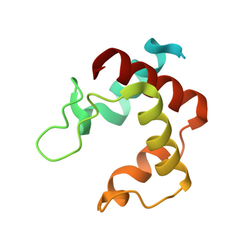
    An interplay between the structural and physicochemical properties of the monoheme cytochromes of type c has been extensively studied. However new proteins belonging to this diverse family continue to reveal some novel and unique features. Here, we present the 1.15 Å structure of the low-potential cytochrome c 546/556 from the bacterium Thioalkalivibrio paradoxus ARh1, which exhibits the prominent splitting of the Q bands in UV-visible spectra even at room temperature. The data obtained suggest that two conformations of the propionate 7 of the heme are responsible for the splitting of the Q bands. We propose that the degree of the splitting of the Q bands is correlated with the conformational lability of the heme propionates.


  • Organizational Affiliation
    • Federal Research Centre «Fundamentals of Biotechnology» of the Russian Academy of Sciences, Leninsky prospect, 33, build. 2, Moscow, 119071, Russian Federation. Electronic address: l.varfolomeeva@fbras.ru.

Macromolecules
Find similar proteins by:  (by identity cutoff)  |  3D Structure
Entity ID: 1
MoleculeChains Sequence LengthOrganismDetailsImage
Cytochrome C
A, B
105Thioalkalivibrio paradoxus ARh 1Mutation(s): 0 
UniProt
Find proteins for W0DLP1 (Thioalkalivibrio paradoxus ARh 1)
Explore W0DLP1 
Go to UniProtKB:  W0DLP1
Entity Groups  
Sequence Clusters30% Identity50% Identity70% Identity90% Identity95% Identity100% Identity
UniProt GroupW0DLP1
Sequence Annotations
Expand
  • Reference Sequence
Experimental Data & Validation

Experimental Data

  • Method: X-RAY DIFFRACTION
  • Resolution: 1.15 Å
  • R-Value Free:  0.144 (Depositor), 0.140 (DCC) 
  • R-Value Work:  0.114 (Depositor), 0.110 (DCC) 
  • R-Value Observed: 0.116 (Depositor) 
Space Group: P 21 21 21
Unit Cell:
Length ( Å )Angle ( ˚ )
a = 36.416α = 90
b = 37.601β = 90
c = 97.117γ = 90
Software Package:
Software NamePurpose
REFMACrefinement
Aimlessdata scaling
CRANK2phasing
DIALSdata reduction

Structure Validation

View Full Validation Report


Ligand Structure Quality Assessment 


Entry History & Funding Information

Deposition Data


Funding OrganizationLocationGrant Number
Not funded--

Revision History  (Full details and data files)

  • Version 1.0: 2024-04-24
    Type: Initial release
  • Version 1.1: 2024-10-23
    Changes: Structure summary
  • Version 1.2: 2025-07-23
    Changes: Database references
  • Version 1.3: 2025-07-30
    Changes: Database references