


structure of IL-4Ra in complex with an nanobody 4E9
Song, G., Qiu, W.To be published.
Experimental Data Snapshot
Starting Model: experimental
View more details
Entity ID: 1 | |||||
|---|---|---|---|---|---|
| Molecule | Chains | Sequence Length | Organism | Details | Image |
| Interleukin-4 receptor subunit alpha | A [auth B] | 214 | Homo sapiens | Mutation(s): 1 Gene Names: IL4R, IL4RA, 582J2.1 | 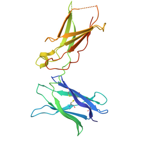 |
UniProt & NIH Common Fund Data Resources | |||||
Find proteins for P24394 (Homo sapiens) Explore P24394 Go to UniProtKB: P24394 | |||||
PHAROS: P24394 GTEx: ENSG00000077238 | |||||
Entity Groups | |||||
| Sequence Clusters | 30% Identity50% Identity70% Identity90% Identity95% Identity100% Identity | ||||
| UniProt Group | P24394 | ||||
Glycosylation | |||||
| Glycosylation Sites: 2 | Go to GlyGen: P24394-1 | ||||
Sequence AnnotationsExpand | |||||
| |||||
Entity ID: 2 | |||||
|---|---|---|---|---|---|
| Molecule | Chains | Sequence Length | Organism | Details | Image |
| nanobody dAb1 | B [auth A] | 124 | Vicugna pacos | Mutation(s): 0 | 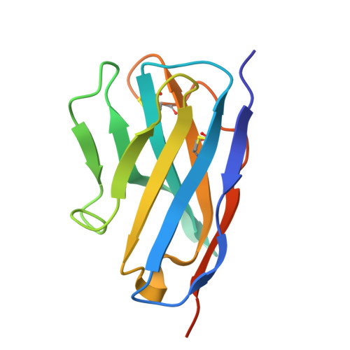 |
Entity Groups | |||||
| Sequence Clusters | 30% Identity50% Identity70% Identity90% Identity95% Identity100% Identity | ||||
Sequence AnnotationsExpand | |||||
| |||||
| Ligands 1 Unique | |||||
|---|---|---|---|---|---|
| ID | Chains | Name / Formula / InChI Key | 2D Diagram | 3D Interactions | |
| NAG Query on NAG | D [auth B] | 2-acetamido-2-deoxy-beta-D-glucopyranose C8 H15 N O6 OVRNDRQMDRJTHS-FMDGEEDCSA-N |  | ||
| Length ( Å ) | Angle ( ˚ ) |
|---|---|
| a = 142.936 | α = 90 |
| b = 142.936 | β = 90 |
| c = 149.087 | γ = 120 |
| Software Name | Purpose |
|---|---|
| PHENIX | refinement |
| Coot | model building |
| XDS | data scaling |
| Funding Organization | Location | Grant Number |
|---|---|---|
| Not funded | -- |


