9IYK | pdb_00009iyk

Crystal structure of hSOD1 in C121 space group


Experimental Data Snapshot

  • Method: X-RAY DIFFRACTION
  • Resolution: 2.34 Å
  • R-Value Free: 
    0.263 (Depositor), 0.260 (DCC) 
  • R-Value Work: 
    0.198 (Depositor), 0.200 (DCC) 
  • R-Value Observed: 
    0.201 (Depositor) 

Starting Model: experimental
View more details

wwPDB Validation   3D Report Full Report


Ligand Structure Quality Assessment 


This is version 1.3 of the entry. See complete history


Literature

Structural Insights into the Dynamics of Water in SOD1 Catalysis and Drug Interactions.

Yapici, I.Tokur, A.G.Sever, B.Ciftci, H.Basak, A.N.DeMirci, H.

(2025) Int J Mol Sci 26

  • DOI: https://doi.org/10.3390/ijms26094228
  • Primary Citation of Related Structures:  
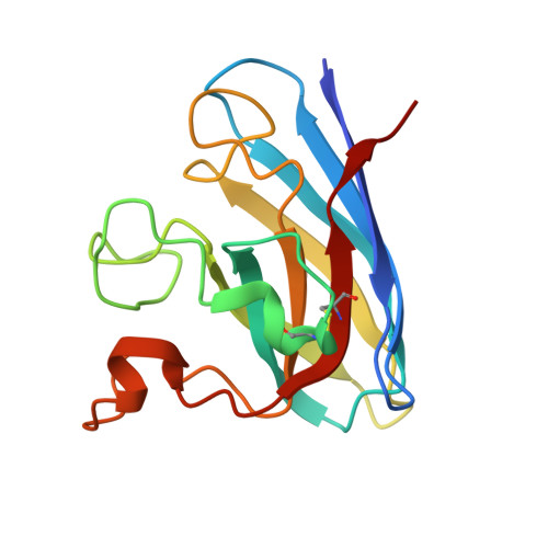
    9IYK

  • PubMed Abstract: 

    Superoxide dismutase 1 (SOD1) is a crucial enzyme that protects cells from oxidative damage by converting superoxide radicals into H 2 O 2 and O 2 . This detoxification process, essential for cellular homeostasis, relies on a precisely orchestrated catalytic mechanism involving the copper cation, while the zinc cation contributes to the structural integrity of the enzyme. This study presents the 2.3 Å crystal structure of human SOD1 (PDB ID: 9IYK), revealing an assembly of six homodimers and twelve distinct active sites. The water molecules form a complex hydrogen-bonding network that drives proton transfer and sustains active site dynamics. Our structure also uncovers subtle conformational changes that highlight the intrinsic flexibility of SOD1, which is essential for its function. Additionally, we observe how these dynamic structural features may be linked to pathological mutations associated with amyotrophic lateral sclerosis (ALS). By advancing our understanding of hSOD1's mechanistic intricacies and the influence of water coordination, this study offers valuable insights for developing therapeutic strategies targeting ALS. Our structure's unique conformations and active site interactions illuminate new facets of hSOD1 function, underscoring the critical role of structural dynamics in enzyme catalysis. Moreover, we conducted a molecular docking analysis using SOD1 for potential radical scavengers and Abelson non-receptor tyrosine kinase (c-Abl, Abl1) inhibitors targeting misfolded SOD1 aggregation along with oxidative stress and apoptosis, respectively. The results showed that CHEMBL1075867, a free radical scavenger derivative, showed the most promising docking results and interactions at the binding site of hSOD1, highlighting its promising role for further studies against SOD1-mediated ALS.


  • Organizational Affiliation
    • Department of Molecular Biology and Genetics, Koc University, Istanbul 34450, Türkiye.

Macromolecules
Find similar proteins by:  (by identity cutoff)  |  3D Structure
Entity ID: 1
MoleculeChains Sequence LengthOrganismDetailsImage
Superoxide dismutase [Cu-Zn]156Homo sapiensMutation(s): 0 
Gene Names: SOD1
EC: 1.15.1.1
UniProt & NIH Common Fund Data Resources
Find proteins for P00441 (Homo sapiens)
Explore P00441 
Go to UniProtKB:  P00441
PHAROS:  P00441
GTEx:  ENSG00000142168 
Entity Groups  
Sequence Clusters30% Identity50% Identity70% Identity90% Identity95% Identity100% Identity
UniProt GroupP00441
Sequence Annotations
Expand
  • Reference Sequence
Small Molecules
Ligands 4 Unique
IDChains Name / Formula / InChI Key2D Diagram3D Interactions
SO4 (Subject of Investigation/LOI)
Query on SO4

Download Ideal Coordinates CCD File 
CA [auth E],
MA [auth I],
Q [auth A],
V [auth C]
SULFATE ION
O4 S
QAOWNCQODCNURD-UHFFFAOYSA-L
ZN (Subject of Investigation/LOI)
Query on ZN

Download Ideal Coordinates CCD File 
BA [auth E]
EA [auth D]
GA [auth B]
JA [auth G]
LA [auth I]
BA [auth E],
EA [auth D],
GA [auth B],
JA [auth G],
LA [auth I],
N [auth A],
O [auth A],
OA [auth L],
QA [auth J],
SA [auth H],
T [auth C],
X [auth K],
Z [auth F]
ZINC ION
Zn
PTFCDOFLOPIGGS-UHFFFAOYSA-N
CU (Subject of Investigation/LOI)
Query on CU

Download Ideal Coordinates CCD File 
AA [auth E]
DA [auth D]
FA [auth B]
IA [auth G]
KA [auth I]
AA [auth E],
DA [auth D],
FA [auth B],
IA [auth G],
KA [auth I],
M [auth A],
NA [auth L],
PA [auth J],
RA [auth H],
S [auth C],
W [auth K],
Y [auth F]
COPPER (II) ION
Cu
JPVYNHNXODAKFH-UHFFFAOYSA-N
ACT (Subject of Investigation/LOI)
Query on ACT

Download Ideal Coordinates CCD File 
HA [auth B],
P [auth A],
R [auth C],
U [auth C]
ACETATE ION
C2 H3 O2
QTBSBXVTEAMEQO-UHFFFAOYSA-M
Experimental Data & Validation

Experimental Data

  • Method: X-RAY DIFFRACTION
  • Resolution: 2.34 Å
  • R-Value Free:  0.263 (Depositor), 0.260 (DCC) 
  • R-Value Work:  0.198 (Depositor), 0.200 (DCC) 
  • R-Value Observed: 0.201 (Depositor) 
Space Group: C 1 2 1
Unit Cell:
Length ( Å )Angle ( ˚ )
a = 178.19α = 90
b = 138.25β = 129.2
c = 112.93γ = 90
Software Package:
Software NamePurpose
PHENIXrefinement
xia2data reduction
xia2data scaling
PHENIXphasing

Structure Validation

View Full Validation Report


Ligand Structure Quality Assessment 


Entry History & Funding Information

Deposition Data


Funding OrganizationLocationGrant Number
Other governmentTurkey122Z429

Revision History  (Full details and data files)

  • Version 1.0: 2024-08-14
    Type: Initial release
  • Version 1.1: 2024-10-16
    Changes: Structure summary
  • Version 1.2: 2025-05-07
    Changes: Database references
  • Version 1.3: 2025-05-28
    Changes: Database references