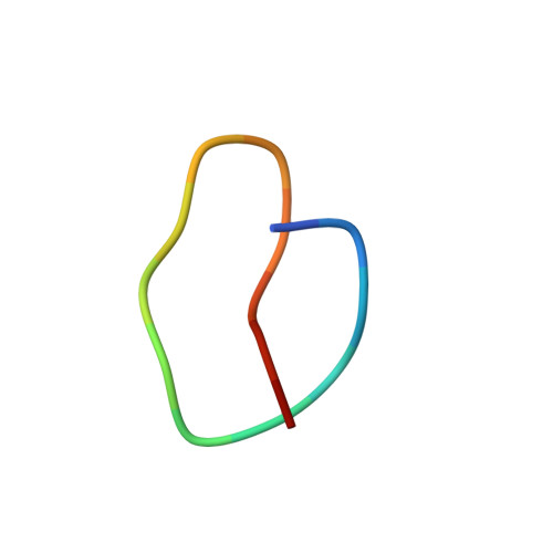
9KDF | pdb_00009kdf

CryoEM structure of Calcineurin-fusion Human endothelin receptor type-B in complex with RES-701-3


Experimental Data Snapshot

  • Method: ELECTRON MICROSCOPY
  • Resolution: 3.30 Å
  • Aggregation State: PARTICLE 
  • Reconstruction Method: SINGLE PARTICLE 

wwPDB Validation   3D Report Full Report


This is version 1.1 of the entry. See complete history


Literature

Structure of a lasso peptide bound ET B receptor provides insights into the mechanism of GPCR inverse agonism.

Shihoya, W.Akasaka, H.Jordan, P.A.Lechner, A.Okada, B.K.Costa Machado da Cruz, G.Sano, F.K.Tanaka, T.Kawahara, R.Chaudhari, R.Masamune, H.Burk, M.J.Nureki, O.

(2025) Nat Commun 16: 3446-3446

  • DOI: https://doi.org/10.1038/s41467-025-57960-x
  • Primary Citation of Related Structures:  
    9KDF, 9KDG

  • PubMed Abstract: 

    Lasso peptides exhibit a unique lariat-like knotted structure imparting exceptional stability and thus show promise as therapeutic agents that target cell-surface receptors. One such receptor is the human endothelin type B receptor (ET B ), which is implicated in challenging cancers with poor immunotherapy responsiveness. The Streptomyces-derived lasso peptide, RES-701-3, is a selective inhibitor for ET B and a compelling candidate for therapeutic development. However, meager production from a genetically recalcitrant host has limited further structure-activity relationship studies of this potent inhibitor. Here, we report cryo-electron microscopy structures of ET B receptor in both its apo form and complex with RES-701-3, facilitated by a calcineurin-fusion strategy. Hydrophobic interactions between RES-701-3 and the transmembrane region of the receptor, especially involving two tryptophan residues, play a crucial role in RES-701-3 binding. Furthermore, RES-701-3 prevents conformational changes associated with G-protein coupling, explaining its inverse agonist activity. A comparative analysis with other lasso peptides and their target proteins highlights the potential of lasso peptides as precise drug candidates for G-protein-coupled receptors. This structural insight into RES-701-3 binding to ET B receptor offers valuable information for the development of novel therapeutics targeting this receptor and provides a broader understanding of lasso peptide interactions with human cell-surface receptors.


  • Organizational Affiliation
    • Department of Biological Sciences, Graduate School of Science, The University of Tokyo, Bunkyo-Ku, Tokyo, Japan. wtrshh9@gmail.com.

Macromolecules
Find similar proteins by:  (by identity cutoff)  |  3D Structure
Entity ID: 1
MoleculeChains Sequence LengthOrganismDetailsImage
Calcineurin-fusion endothelin receptor type-BA [auth R]908Homo sapiensMutation(s): 0 
Entity Groups  
Sequence Clusters30% Identity50% Identity70% Identity90% Identity95% Identity100% Identity
Sequence Annotations
Expand
  • Reference Sequence
Find similar proteins by:  (by identity cutoff)  |  3D Structure
Entity ID: 2
MoleculeChains Sequence LengthOrganismDetailsImage
Peptidyl-prolyl cis-trans isomerase FKBP1BB [auth F]116Homo sapiensMutation(s): 0 
Gene Names: FKBP1BFKBP12.6FKBP1LFKBP9OTK4
EC: 5.2.1.8
UniProt & NIH Common Fund Data Resources
Find proteins for P68106 (Homo sapiens)
Explore P68106 
Go to UniProtKB:  P68106
PHAROS:  P68106
GTEx:  ENSG00000119782 
Entity Groups  
Sequence Clusters30% Identity50% Identity70% Identity90% Identity95% Identity100% Identity
UniProt GroupP68106
Sequence Annotations
Expand
  • Reference Sequence

Find similar proteins by:  Sequence   |   3D Structure  

Entity ID: 3
MoleculeChains Sequence LengthOrganismDetailsImage
RES-701-3C [auth A]16Streptomyces venezuelaeMutation(s): 0 
Entity Groups  
Sequence Clusters30% Identity50% Identity70% Identity90% Identity95% Identity100% Identity
Sequence Annotations
Expand
  • Reference Sequence
Small Molecules
Ligands 4 Unique
IDChains Name / Formula / InChI Key2D Diagram3D Interactions
FK5
Query on FK5

Download Ideal Coordinates CCD File 
J [auth F]8-DEETHYL-8-[BUT-3-ENYL]-ASCOMYCIN
C44 H69 N O12
QJJXYPPXXYFBGM-LFZNUXCKSA-N
ZN
Query on ZN

Download Ideal Coordinates CCD File 
I [auth R]ZINC ION
Zn
PTFCDOFLOPIGGS-UHFFFAOYSA-N
FE
Query on FE

Download Ideal Coordinates CCD File 
H [auth R]FE (III) ION
Fe
VTLYFUHAOXGGBS-UHFFFAOYSA-N
CA
Query on CA

Download Ideal Coordinates CCD File 
D [auth R],
E [auth R],
F [auth R],
G [auth R]
CALCIUM ION
Ca
BHPQYMZQTOCNFJ-UHFFFAOYSA-N
Experimental Data & Validation

Experimental Data

  • Method: ELECTRON MICROSCOPY
  • Resolution: 3.30 Å
  • Aggregation State: PARTICLE 
  • Reconstruction Method: SINGLE PARTICLE 
EM Software:
TaskSoftware PackageVersion
MODEL REFINEMENTPHENIX

Structure Validation

View Full Validation Report


Entry History & Funding Information

Deposition Data


Funding OrganizationLocationGrant Number
Japan Society for the Promotion of Science (JSPS)Japan21H05037

Revision History  (Full details and data files)

  • Version 1.0: 2025-05-07
    Type: Initial release
  • Version 1.1: 2025-07-02
    Changes: Data collection