9B81 | pdb_00009b81

Crystal structure of wild type IDH1 bound to compound 4


Experimental Data Snapshot

  • Method: X-RAY DIFFRACTION
  • Resolution: 2.56 Å
  • R-Value Free: 
    0.233 (Depositor), 0.235 (DCC) 
  • R-Value Work: 
    0.201 (Depositor), 0.202 (DCC) 
  • R-Value Observed: 
    0.203 (Depositor) 

Starting Model: experimental
View more details

wwPDB Validation   3D Report Full Report


Ligand Structure Quality Assessment 


This is version 1.0 of the entry. See complete history


Literature

Biostructural, biochemical and biophysical studies of mutant IDH1.

McCoy, M.A.Lu, J.Richard Miller, F.Soisson, S.M.Lam, M.H.Fischer, C.

(2024) Nat Commun 15: 7877-7877

  • DOI: https://doi.org/10.1038/s41467-024-51692-0
  • Primary Citation of Related Structures:  
    8T7D, 8T7N, 8T7O, 9B81

  • PubMed Abstract: 

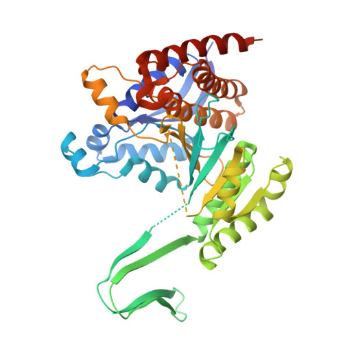
    We report bio-structural, bio-chemical and bio-physical evidence demonstrating how small molecules can bind to both wild-type and mutant IDH1, but only inhibit the enzymatic activity of the mutant isoform. Enabled through x-ray crystallography, we characterized a series of small molecule inhibitors that bound to mutant IDH1 differently than the marketed inhibitor Ivosidenib, for which we have determined the x-ray crystal structure. Across the industry several mutant IDH1 inhibitor chemotypes bind to this allosteric IDH1 pocket and selectively inhibit the mutant enzyme. Detailed characterization by a variety of biophysical techniques and NMR studies led us to propose how compounds binding in the allosteric IDH1 R132H pocket inhibit the production of 2-Hydroxy glutarate.


  • Organizational Affiliation

    MRL, Merck & Co., Inc., Rahway, NJ, USA. mark.mccoy@merck.com.


Macromolecules
Find similar proteins by:  (by identity cutoff)  |  3D Structure
Entity ID: 1
MoleculeChains Sequence LengthOrganismDetailsImage
Isocitrate dehydrogenase [NADP] cytoplasmic
A, B
424Homo sapiensMutation(s): 1 
Gene Names: IDH1PICD
EC: 1.1.1.42
UniProt & NIH Common Fund Data Resources
Find proteins for O75874 (Homo sapiens)
Explore O75874 
Go to UniProtKB:  O75874
PHAROS:  O75874
GTEx:  ENSG00000138413 
Entity Groups  
Sequence Clusters30% Identity50% Identity70% Identity90% Identity95% Identity100% Identity
UniProt GroupO75874
Sequence Annotations
Expand
  • Reference Sequence
Small Molecules
Ligands 3 Unique
IDChains Name / Formula / InChI Key2D Diagram3D Interactions
NAP
Query on NAP

Download Ideal Coordinates CCD File 
D [auth A],
G [auth B]
NADP NICOTINAMIDE-ADENINE-DINUCLEOTIDE PHOSPHATE
C21 H28 N7 O17 P3
XJLXINKUBYWONI-NNYOXOHSSA-N
A1AI4 (Subject of Investigation/LOI)
Query on A1AI4

Download Ideal Coordinates CCD File 
C [auth A],
F [auth B]
2-methyl-2-(6-{4-[(2S)-1,1,1-trifluoro-2-hydroxypropan-2-yl]benzoyl}-6,11-dihydro-5H-pyrido[2,3-b][1,5]benzodiazepin-8-yl)propanenitrile
C26 H23 F3 N4 O2
ARTKCJPBKAKQRY-VWLOTQADSA-N
GOL
Query on GOL

Download Ideal Coordinates CCD File 
E [auth A]GLYCEROL
C3 H8 O3
PEDCQBHIVMGVHV-UHFFFAOYSA-N
Experimental Data & Validation

Experimental Data

  • Method: X-RAY DIFFRACTION
  • Resolution: 2.56 Å
  • R-Value Free:  0.233 (Depositor), 0.235 (DCC) 
  • R-Value Work:  0.201 (Depositor), 0.202 (DCC) 
  • R-Value Observed: 0.203 (Depositor) 
Space Group: P 43 21 2
Unit Cell:
Length ( Å )Angle ( ˚ )
a = 82.79α = 90
b = 82.79β = 90
c = 302.16γ = 90
Software Package:
Software NamePurpose
BUSTERrefinement
XDSdata reduction
XDSdata scaling
PHASERphasing

Structure Validation

View Full Validation Report


Ligand Structure Quality Assessment 


Entry History & Funding Information

Deposition Data


Funding OrganizationLocationGrant Number
Not funded--

Revision History  (Full details and data files)

  • Version 1.0: 2025-03-26
    Type: Initial release