9BPC | pdb_00009bpc

Cryo-EM structure of P2X3 receptor in complex with camlipixant


Experimental Data Snapshot

  • Method: ELECTRON MICROSCOPY
  • Resolution: 3.44 Å
  • Aggregation State: PARTICLE 
  • Reconstruction Method: SINGLE PARTICLE 

Starting Model: in silico
View more details

wwPDB Validation   3D Report Full Report


This is version 1.0 of the entry. See complete history


Literature

Mechanistic insights into the selective targeting of P2X3 receptor by camlipixant antagonist.

Thach, T.Dhanabalan, K.Nandekar, P.P.Stauffer, S.Heisler, I.Alvarado, S.Snyder, J.Subramanian, R.

(2025) J Biological Chem 301: 108109-108109

  • DOI: https://doi.org/10.1016/j.jbc.2024.108109
  • Primary Citation of Related Structures:  
    9BPC, 9BPD

  • PubMed Abstract: 

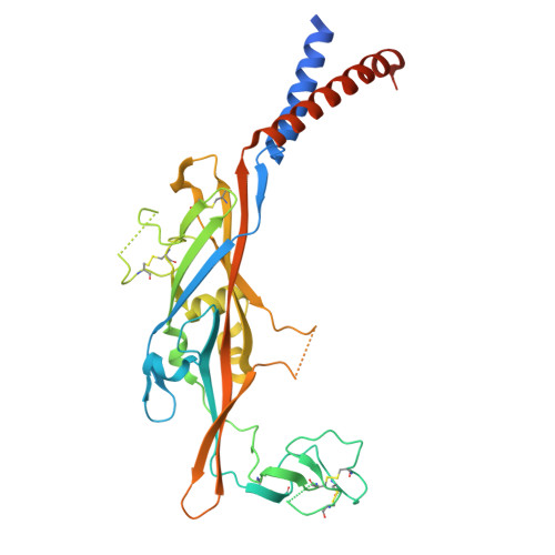
    ATP-activated P2X3 receptors play a pivotal role in chronic cough, affecting more than 10% of the population. Despite the challenges posed by the highly conserved structure of P2X receptors, efforts to develop selective drugs targeting P2X3 have led to the development of camlipixant, a potent, selective P2X3 antagonist. However, the mechanisms of receptor desensitization, ion permeation, and structural basis of camlipixant binding to P2X3 remain unclear. Here, we report a cryo-EM structure of camlipixant-bound P2X3, revealing a previously undiscovered selective drug-binding site in the receptor. Our findings also demonstrate that conformational changes in the upper body domain, including the turret and camlipixant-binding pocket, play a critical role: turret opening facilitates P2X3 channel closure to a radius of 0.7 Å, hindering cation transfer, whereas turret closure leads to channel opening. Structural and functional studies combined with molecular dynamics simulations provide a comprehensive understanding of camlipixant's selective inhibition of P2X3, offering a foundation for future drug development targeting this receptor.


  • Organizational Affiliation
    • Department of Biological Sciences, Purdue University, West Lafayette, Indiana, USA. Electronic address: ttthach@purdue.edu.

Macromolecules
Find similar proteins by:  (by identity cutoff)  |  3D Structure
Entity ID: 1
MoleculeChains Sequence LengthOrganismDetailsImage
P2X purinoceptor
A, B, C
361Canis lupusMutation(s): 0 
Gene Names: P2RX3
UniProt
Find proteins for A0A8I3S575 (Canis lupus familiaris)
Explore A0A8I3S575 
Go to UniProtKB:  A0A8I3S575
Entity Groups  
Sequence Clusters30% Identity50% Identity70% Identity90% Identity95% Identity100% Identity
UniProt GroupA0A8I3S575
Glycosylation
Glycosylation Sites: 2
Sequence Annotations
Expand
  • Reference Sequence
Oligosaccharides

Help

Entity ID: 2
MoleculeChains Length2D Diagram Glycosylation3D Interactions
2-acetamido-2-deoxy-beta-D-glucopyranose-(1-4)-2-acetamido-2-deoxy-beta-D-glucopyranose
D, E, F
2N-Glycosylation
Glycosylation Resources
GlyTouCan:  G42666HT
GlyCosmos:  G42666HT
GlyGen:  G42666HT
Experimental Data & Validation

Experimental Data

  • Method: ELECTRON MICROSCOPY
  • Resolution: 3.44 Å
  • Aggregation State: PARTICLE 
  • Reconstruction Method: SINGLE PARTICLE 
EM Software:
TaskSoftware PackageVersion
RECONSTRUCTIONcryoSPARC4.4.1
MODEL REFINEMENTPHENIX1.21.1_5286:

Structure Validation

View Full Validation Report


Entry History & Funding Information

Deposition Data


Funding OrganizationLocationGrant Number
Not funded--

Revision History  (Full details and data files)

  • Version 1.0: 2025-03-19
    Type: Initial release