9BWC | pdb_00009bwc

Homomeric alpha3 glycine receptor in the presence of 0.1 mM glycine at pH 6.4 in an apo state


Experimental Data Snapshot

  • Method: ELECTRON MICROSCOPY
  • Resolution: 2.19 Å
  • Aggregation State: PARTICLE 
  • Reconstruction Method: SINGLE PARTICLE 

wwPDB Validation   3D Report Full Report


This is version 1.2 of the entry. See complete history


Literature

Mechanisms underlying modulation of human GlyR alpha 3 by Zn 2+ and pH.

Kindig, K.Gibbs, E.Seiferth, D.Biggin, P.C.Chakrapani, S.

(2024) Sci Adv 10: eadr5920-eadr5920

  • DOI: https://doi.org/10.1126/sciadv.adr5920
  • Primary Citation of Related Structures:  
    9BU2, 9BU3, 9BVH, 9BVJ, 9BWB, 9BWC, 9BWE, 9BWG, 9BWJ, 9BZP

  • PubMed Abstract: 

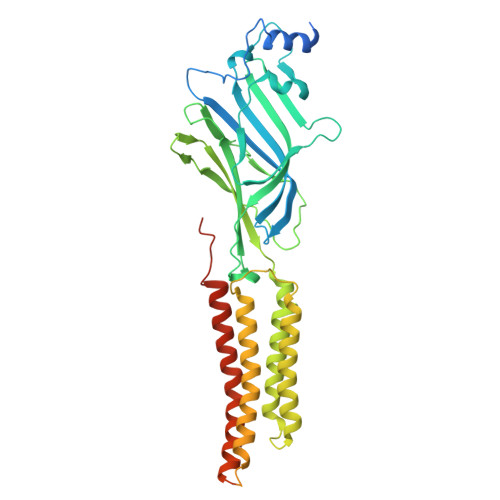
    Glycine receptors (GlyRs) regulate motor control and pain processing in the central nervous system through inhibitory synaptic signaling. The subtype GlyRα3 expressed in nociceptive sensory neurons of the spinal dorsal horn is a key regulator of physiological pain perception. Disruption of spinal glycinergic inhibition is associated with chronic inflammatory pain states, making GlyRα3 an attractive target for pain treatment. GlyRα3 activity is modulated by numerous endogenous and exogenous ligands that consequently affect pain sensitization. To understand the mechanism of two such endogenous modulators, Zn 2+ and protons, we have used cryo-electron microscopy to determine structures of full-length human GlyRα3 in various functional states. Whereas acidic pH reduces peak glycine response, Zn 2+ displays biphasic modulation in a concentration-dependent manner. Our findings reveal the effector sites and also capture intermediate conformations in the gating cycle. Combined with molecular dynamics simulations and electrophysiology, this work provides important insights into GlyRα3 activation and regulation.


  • Organizational Affiliation
    • Department of Physiology and Biophysics, School of Medicine, Case Western Reserve University, Cleveland, OH 44106-4970, USA.

Macromolecules
Find similar proteins by:  (by identity cutoff)  |  3D Structure
Entity ID: 1
MoleculeChains Sequence LengthOrganismDetailsImage
Glycine receptor subunit alpha-3
A, B, C, D, E
478Homo sapiensMutation(s): 0 
Gene Names: GLRA3
Membrane Entity: Yes 
UniProt & NIH Common Fund Data Resources
Find proteins for O75311 (Homo sapiens)
Explore O75311 
Go to UniProtKB:  O75311
PHAROS:  O75311
GTEx:  ENSG00000145451 
Entity Groups  
Sequence Clusters30% Identity50% Identity70% Identity90% Identity95% Identity100% Identity
UniProt GroupO75311
Glycosylation
Glycosylation Sites: 1Go to GlyGen: O75311-1
Sequence Annotations
Expand
  • Reference Sequence
Oligosaccharides

Help

Entity ID: 2
MoleculeChains Length2D Diagram Glycosylation3D Interactions
2-acetamido-2-deoxy-beta-D-glucopyranose-(1-4)-2-acetamido-2-deoxy-beta-D-glucopyranose
F, G, H, I, J
2N-Glycosylation
Glycosylation Resources
GlyTouCan:  G42666HT
GlyCosmos:  G42666HT
GlyGen:  G42666HT
Small Molecules
Ligands 3 Unique
IDChains Name / Formula / InChI Key2D Diagram3D Interactions
PIO
Query on PIO

Download Ideal Coordinates CCD File 
AA [auth C],
HA [auth D],
L [auth A],
SA [auth E],
T [auth B]
[(2R)-2-octanoyloxy-3-[oxidanyl-[(1R,2R,3S,4R,5R,6S)-2,3,6-tris(oxidanyl)-4,5-diphosphonooxy-cyclohexyl]oxy-phosphoryl]oxy-propyl] octanoate
C25 H49 O19 P3
XLNCEHRXXWQMPK-MJUMVPIBSA-N
PX4
Query on PX4

Download Ideal Coordinates CCD File 
BA [auth C]
CA [auth C]
DA [auth C]
EA [auth C]
GA [auth D]
BA [auth C],
CA [auth C],
DA [auth C],
EA [auth C],
GA [auth D],
IA [auth D],
JA [auth D],
K [auth A],
KA [auth D],
LA [auth D],
M [auth A],
MA [auth E],
N [auth A],
NA [auth E],
O [auth A],
OA [auth E],
P [auth A],
PA [auth E],
RA [auth E],
S [auth B],
U [auth B],
V [auth B],
W [auth B],
X [auth B],
Z [auth C]
1,2-DIMYRISTOYL-SN-GLYCERO-3-PHOSPHOCHOLINE
C36 H73 N O8 P
CITHEXJVPOWHKC-UUWRZZSWSA-O
GLY (Subject of Investigation/LOI)
Query on GLY

Download Ideal Coordinates CCD File 
FA [auth D],
Q [auth A],
QA [auth E],
R [auth B],
Y [auth C]
GLYCINE
C2 H5 N O2
DHMQDGOQFOQNFH-UHFFFAOYSA-N
Experimental Data & Validation

Experimental Data

  • Method: ELECTRON MICROSCOPY
  • Resolution: 2.19 Å
  • Aggregation State: PARTICLE 
  • Reconstruction Method: SINGLE PARTICLE 

Structure Validation

View Full Validation Report


Entry History & Funding Information

Deposition Data


Funding OrganizationLocationGrant Number
National Institutes of Health/National Institute of General Medical Sciences (NIH/NIGMS)United StatesR01GM131216
National Institutes of Health/National Institute of General Medical Sciences (NIH/NIGMS)United StatesR35GM134896
National Institutes of Health/National Institute of General Medical Sciences (NIH/NIGMS)United States3R01GM108921-03S1
National Institutes of Health/National Institute of General Medical Sciences (NIH/NIGMS)United StatesR01GM108921-5S1
National Institutes of Health/National Institute of General Medical Sciences (NIH/NIGMS)United States3R01GM131216-1S1
National Institutes of Health/Office of the DirectorUnited States1S10OD032437

Revision History  (Full details and data files)

  • Version 1.0: 2024-11-13
    Type: Initial release
  • Version 1.1: 2024-11-27
    Changes: Data collection
  • Version 1.2: 2025-05-28
    Changes: Data collection, Database references