9C4D | pdb_00009c4d

The structure of 4 MntR homodimers bound to the promoter sequence of mnep


Experimental Data Snapshot

  • Method: ELECTRON MICROSCOPY
  • Resolution: 4.17 Å
  • Aggregation State: PARTICLE 
  • Reconstruction Method: SINGLE PARTICLE 

wwPDB Validation   3D Report Full Report


This is version 1.3 of the entry. See complete history


Literature

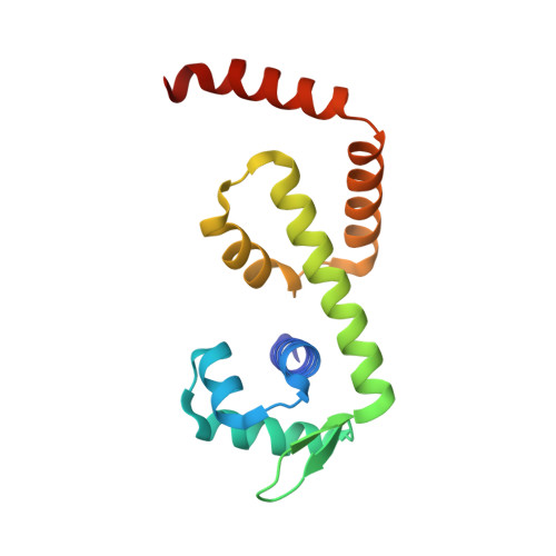
Structural basis for transcription activation through cooperative recruitment of MntR.

Shi, H.Fu, Y.Kodyte, V.Andreas, A.Sachla, A.J.Miller, K.Shrestha, R.Helmann, J.D.Glasfeld, A.Ahuja, S.

(2025) Nat Commun 16: 2204-2204

  • DOI: https://doi.org/10.1038/s41467-025-57412-6
  • Primary Citation of Related Structures:  
    9C4C, 9C4D

  • PubMed Abstract: 

    Bacillus subtilis MntR is a dual regulatory protein that responds to heightened Mn 2+ availability in the cell by both repressing the expression of uptake transporters and activating the expression of efflux proteins. Recent work indicates that, in its role as an activator, MntR binds several sites upstream of the genes encoding Mn 2+ exporters, leading to a cooperative response to manganese. Here, we use cryo-EM to explore the molecular basis of gene activation by MntR and report a structure of four MntR dimers bound to four 18-base pair sites across an 84-base pair regulatory region of the mneP promoter. Our structures, along with solution studies including mass photometry and in vivo transcription assays, reveal that MntR dimers employ polar and non-polar contacts to bind cooperatively to an array of low-affinity DNA-binding sites. These results reveal the molecular basis for cooperativity in the activation of manganese efflux.


  • Organizational Affiliation
    • Department of Chemistry, Reed College, Portland, Oregon, USA.

Macromolecules

Find similar proteins by:  (by identity cutoff)  |  3D Structure
Entity ID: 3
MoleculeChains Sequence LengthOrganismDetailsImage
HTH-type transcriptional regulator MntR
C, D, E, F, G
C, D, E, F, G, H, I, J
142Bacillus subtilisMutation(s): 0 
Gene Names: mntRyqhNBSU24520
UniProt
Find proteins for P54512 (Bacillus subtilis (strain 168))
Explore P54512 
Go to UniProtKB:  P54512
Entity Groups  
Sequence Clusters30% Identity50% Identity70% Identity90% Identity95% Identity100% Identity
UniProt GroupP54512
Sequence Annotations
Expand
  • Reference Sequence
Find similar nucleic acids by:  (by identity cutoff)  |  3D Structure
Entity ID: 1
MoleculeChains LengthOrganismImage
DNA (77-MER)77Bacillus subtilis
Sequence Annotations
Expand
  • Reference Sequence
Find similar nucleic acids by:  (by identity cutoff)  |  3D Structure
Entity ID: 2
MoleculeChains LengthOrganismImage
DNA (77-MER)77Bacillus subtilis
Sequence Annotations
Expand
  • Reference Sequence
Small Molecules
Ligands 1 Unique
IDChains Name / Formula / InChI Key2D Diagram3D Interactions
MN
Query on MN

Download Ideal Coordinates CCD File 
K [auth C]
L [auth C]
M [auth D]
N [auth D]
O [auth E]
K [auth C],
L [auth C],
M [auth D],
N [auth D],
O [auth E],
P [auth E],
Q [auth F],
R [auth F],
S [auth G],
T [auth G],
U [auth H],
V [auth H],
W [auth I],
X [auth I],
Y [auth J],
Z [auth J]
MANGANESE (II) ION
Mn
WAEMQWOKJMHJLA-UHFFFAOYSA-N
Experimental Data & Validation

Experimental Data

  • Method: ELECTRON MICROSCOPY
  • Resolution: 4.17 Å
  • Aggregation State: PARTICLE 
  • Reconstruction Method: SINGLE PARTICLE 
EM Software:
TaskSoftware PackageVersion
MODEL REFINEMENTPHENIX

Structure Validation

View Full Validation Report


Entry History & Funding Information

Deposition Data


Funding OrganizationLocationGrant Number
Not funded--

Revision History  (Full details and data files)

  • Version 1.0: 2024-08-14
    Type: Initial release
  • Version 1.1: 2025-03-12
    Changes: Data collection, Database references, Structure summary
  • Version 1.2: 2025-03-19
    Changes: Data collection, Database references
  • Version 1.3: 2025-05-14
    Changes: Data collection