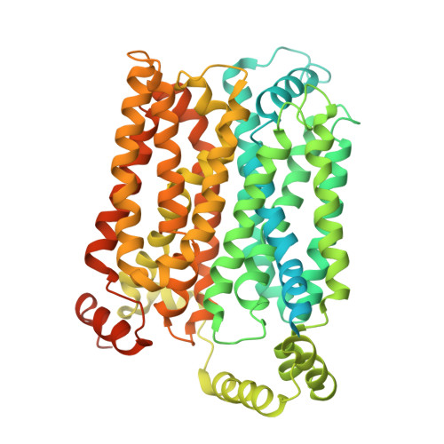
Structural basis of disease mutation and substrate recognition by the human SLC2A9 transporter.
Khandelwal, N.K., Gupta, M., Kumar, P., Balasubramani, S.G., Echeverria, I., Stroud, R.M.(2025) Proc Natl Acad Sci U S A 122: e2418282122-e2418282122
- PubMed: 39937868
- DOI: https://doi.org/10.1073/pnas.2418282122
- Primary Citation of Related Structures:
9CAX, 9CBB - PubMed Abstract:
Urate provides ~50% of the reducing potential in human and primate plasma which is key to detoxifying reactive oxygen by-products of cellular metabolism. Urate is the endpoint of purine metabolism in primates, and its concentration in plasma is a balance between excretion from kidney and intestine, and subsequent reabsorption in and through cells of kidney proximal tubules to maintain a regulated concentration in plasma. SLC2A9 is the primary transporter that returns urate from the basolateral side of kidney tubule cells back to plasma. A shorter splice variant of SLC2A9 is directed to the apical surface where several transporters recapture urate from the tubule back into cells. Too high a concentration in plasma causes hyperuricemia, is linked to gout, and favors kidney stone formation. To understand the molecular basis of uric acid transport and the role of disease-causing mutations in SLC2A9, we determined structures of human SLC2A9 in its apo form, and its urate-bound form by cryo-EM, at resolution of 3.3 Å and 4.1 Å respectively. Both structures are captured in an inward open conformation. Using the inward-facing structure as a template we modeled the outward-facing conformation to understand the alternating access mechanism. Alternative salt bridge pairs on the cytoplasmic side suggest a mechanism that can balance the energetics of the inward open and outward open states. The location of disease-causing mutants suggests their role in impacting function. Our structures elucidate the molecular basis for urate selectivity and transport and provide a platform for future structure-based drug discovery aimed at reducing plasma urate levels in diseases of hyperuricemia and gout.
- Department of Biochemistry and Biophysics, University of California, San Francisco, San Francisco, CA 94143.
Organizational Affiliation: