9CIB | pdb_00009cib

CryoEM Structure of HCA2 DREADD Gi1 in complex with FCH-2296413 (Local Refinement)


Experimental Data Snapshot

  • Method: ELECTRON MICROSCOPY
  • Resolution: 2.62 Å
  • Aggregation State: PARTICLE 
  • Reconstruction Method: SINGLE PARTICLE 

wwPDB Validation   3D Report Full Report


This is version 1.0 of the entry. See complete history


Literature

Structure-guided design of a peripherally restricted chemogenetic system.

Kang, H.J.Krumm, B.E.Tassou, A.Geron, M.DiBerto, J.F.Kapolka, N.J.Gumpper, R.H.Sakamoto, K.Dewran Kocak, D.Olsen, R.H.J.Huang, X.P.Zhang, S.Huang, K.L.Zaidi, S.A.Nguyen, M.T.Jo, M.J.Katritch, V.Fay, J.F.Scherrer, G.Roth, B.L.

(2024) Cell 187: 7433-7449.e20

  • DOI: https://doi.org/10.1016/j.cell.2024.11.001
  • Primary Citation of Related Structures:  
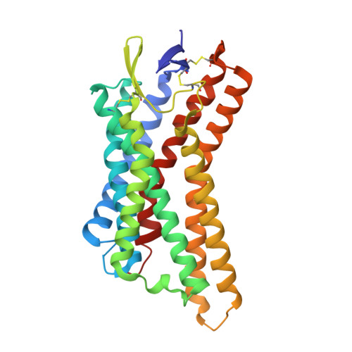
    8UTD, 8UUJ, 9CIB

  • PubMed Abstract: 

    Designer receptors exclusively activated by designer drugs (DREADDs) are chemogenetic tools for remotely controlling cellular signaling, neural activity, behavior, and physiology. Using a structure-guided approach, we provide a peripherally restricted Gi-DREADD, hydroxycarboxylic acid receptor DREADD (HCAD), whose native receptor is minimally expressed in the brain, and a chemical actuator that does not cross the blood-brain barrier (BBB). This was accomplished by combined mutagenesis, analoging via an ultra-large make-on-demand library, structural determination of the designed DREADD receptor via cryoelectron microscopy (cryo-EM), and validation of HCAD function. Expression and activation of HCAD in dorsal root ganglion (DRG) neurons inhibit action potential (AP) firing and reduce both acute and tissue-injury-induced inflammatory pain. The HCAD chemogenetic system expands the possibilities for studying numerous peripheral systems with little adverse effects on the central nervous system (CNS). The structure-guided approach used to generate HCAD also has the potential to accelerate the development of emerging chemogenetic tools for basic and translational sciences.


  • Organizational Affiliation
    • Department of Biotechnology, College of Life Science and Biotechnology, Yonsei University, Seoul, South Korea.

Macromolecules
Find similar proteins by:  (by identity cutoff)  |  3D Structure
Entity ID: 1
MoleculeChains Sequence LengthOrganismDetailsImage
Hydroxycarboxylic acid receptor 2A [auth R]294Mus musculusMutation(s): 2 
Gene Names: Hcar2Gpr109Gpr109aGpr109bNiacr1Pumag
Membrane Entity: Yes 
UniProt & NIH Common Fund Data Resources
Find proteins for Q9EP66 (Mus musculus)
Explore Q9EP66 
Go to UniProtKB:  Q9EP66
IMPC:  MGI:1933383
Entity Groups  
Sequence Clusters30% Identity50% Identity70% Identity90% Identity95% Identity100% Identity
UniProt GroupQ9EP66
Sequence Annotations
Expand
  • Reference Sequence
Small Molecules
Ligands 1 Unique
IDChains Name / Formula / InChI Key2D Diagram3D Interactions
XI9 (Subject of Investigation/LOI)
Query on XI9

Download Ideal Coordinates CCD File 
B [auth R](3M,4aR,5aR)-3-(1H-tetrazol-5-yl)-4,4a,5,5a-tetrahydro-1H-cyclopropa[4,5]cyclopenta[1,2-c]pyrazole
C8 H8 N6
DKSPRNJUDCWTID-QWWZWVQMSA-N
Experimental Data & Validation

Experimental Data

  • Method: ELECTRON MICROSCOPY
  • Resolution: 2.62 Å
  • Aggregation State: PARTICLE 
  • Reconstruction Method: SINGLE PARTICLE 
EM Software:
TaskSoftware PackageVersion
MODEL REFINEMENTPHENIX

Structure Validation

View Full Validation Report


Entry History & Funding Information

Deposition Data


Funding OrganizationLocationGrant Number
National Institutes of Health/National Institute of Mental Health (NIH/NIMH)United States5R01MH112205

Revision History  (Full details and data files)

  • Version 1.0: 2025-05-14
    Type: Initial release