9CYM | pdb_00009cym

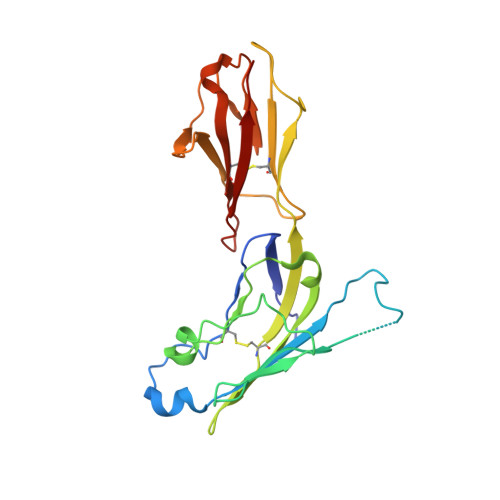
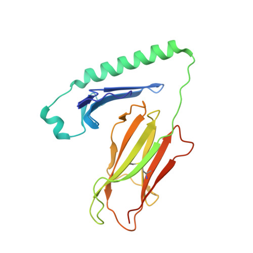
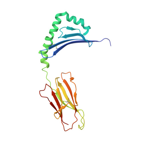
Structure of LAG3 bound to the MHC class II molecule I-A(b)


Experimental Data Snapshot

  • Method: X-RAY DIFFRACTION
  • Resolution: 3.84 Å
  • R-Value Free: 
    0.307 (Depositor), 0.300 (DCC) 
  • R-Value Work: 
    0.260 (Depositor), 0.260 (DCC) 
  • R-Value Observed: 
    0.262 (Depositor) 

Starting Models: experimental
View more details

wwPDB Validation   3D Report Full Report


Ligand Structure Quality Assessment 


This is version 1.2 of the entry. See complete history


Literature

Structural basis for mouse LAG3 interactions with the MHC class II molecule I-A b.

Ming, Q.Antfolk, D.Price, D.A.Manturova, A.Medina, E.Singh, S.Mason, C.Tran, T.H.Smalley, K.S.M.Leung, D.W.Luca, V.C.

(2024) Nat Commun 15: 7513-7513

  • DOI: https://doi.org/10.1038/s41467-024-51930-5
  • Primary Citation of Related Structures:  
    9CYL, 9CYM

  • PubMed Abstract: 

    The immune checkpoint protein, Lymphocyte activation gene-3 (LAG3), binds Major Histocompatibility Complex Class II (MHC-II) and suppresses T cell activation. Despite the recent FDA approval of a LAG3 inhibitor for the treatment of melanoma, how LAG3 engages MHC-II on the cell surface remains poorly understood. Here, we determine the 3.84 Å-resolution structure of mouse LAG3 bound to the MHC-II molecule I-A b , revealing that domain 1 (D1) of LAG3 binds a conserved, membrane-proximal region of MHC-II spanning both the α2 and β2 subdomains. LAG3 dimerization restricts the intermolecular spacing of MHC-II molecules, which may attenuate T cell activation by enforcing suboptimal signaling geometry. The LAG3-MHC-II interface overlaps with the MHC-II-binding site of the T cell coreceptor CD4, implicating disruption of CD4-MHC-II interactions as a mechanism for LAG3 immunosuppressive function. Lastly, antibody epitope analysis indicates that multiple LAG3 inhibitors do not recognize the MHC-II-binding interface of LAG3, suggesting a role for functionally distinct mechanisms of LAG3 antagonism in therapeutic development.


  • Organizational Affiliation

    Moffitt Cancer Center and Research Institute, Department of Immunology, Tampa, FL, 33612, USA.


Macromolecules
Find similar proteins by:  (by identity cutoff)  |  3D Structure
Entity ID: 1
MoleculeChains Sequence LengthOrganismDetailsImage
Secreted lymphocyte activation gene 3 proteinA [auth L]233Mus musculusMutation(s): 0 
Gene Names: Lag3
UniProt
Find proteins for Q61790 (Mus musculus)
Explore Q61790 
Go to UniProtKB:  Q61790
Entity Groups  
Sequence Clusters30% Identity50% Identity70% Identity90% Identity95% Identity100% Identity
UniProt GroupQ61790
Glycosylation
Glycosylation Sites: 2Go to GlyGen: Q61790-1
Sequence Annotations
Expand
  • Reference Sequence
Find similar proteins by:  (by identity cutoff)  |  3D Structure
Entity ID: 2
MoleculeChains Sequence LengthOrganismDetailsImage
H-2 class II histocompatibility antigen, A-B alpha chainB [auth A]195Mus musculusMutation(s): 0 
Gene Names: H2-Aa
UniProt
Find proteins for P14434 (Mus musculus)
Explore P14434 
Go to UniProtKB:  P14434
Entity Groups  
Sequence Clusters30% Identity50% Identity70% Identity90% Identity95% Identity100% Identity
UniProt GroupP14434
Glycosylation
Glycosylation Sites: 1
Sequence Annotations
Expand
  • Reference Sequence

Find similar proteins by:  Sequence   |   3D Structure  

Entity ID: 3
MoleculeChains Sequence LengthOrganismDetailsImage
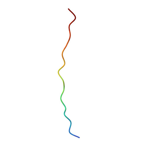
Class-II-associated invariant chain peptideC [auth P]16Homo sapiensMutation(s): 0 
Gene Names: CD74DHLAG
UniProt & NIH Common Fund Data Resources
Find proteins for P04233 (Homo sapiens)
Explore P04233 
Go to UniProtKB:  P04233
PHAROS:  P04233
GTEx:  ENSG00000019582 
Entity Groups  
Sequence Clusters30% Identity50% Identity70% Identity90% Identity95% Identity100% Identity
UniProt GroupP04233
Sequence Annotations
Expand
  • Reference Sequence
Find similar proteins by:  (by identity cutoff)  |  3D Structure
Entity ID: 4
MoleculeChains Sequence LengthOrganismDetailsImage
H-2 class II histocompatibility antigen, A beta chainD [auth B]191Mus musculusMutation(s): 0 
Gene Names: H2-Ab1H2-iabeta
UniProt
Find proteins for P14483 (Mus musculus)
Explore P14483 
Go to UniProtKB:  P14483
Entity Groups  
Sequence Clusters30% Identity50% Identity70% Identity90% Identity95% Identity100% Identity
UniProt GroupP14483
Glycosylation
Glycosylation Sites: 1
Sequence Annotations
Expand
  • Reference Sequence
Experimental Data & Validation

Experimental Data

  • Method: X-RAY DIFFRACTION
  • Resolution: 3.84 Å
  • R-Value Free:  0.307 (Depositor), 0.300 (DCC) 
  • R-Value Work:  0.260 (Depositor), 0.260 (DCC) 
  • R-Value Observed: 0.262 (Depositor) 
Space Group: P 65 2 2
Unit Cell:
Length ( Å )Angle ( ˚ )
a = 187.652α = 90
b = 187.652β = 90
c = 124.057γ = 120
Software Package:
Software NamePurpose
PHENIXrefinement
XSCALEdata scaling
XDSdata reduction
PHASERphasing

Structure Validation

View Full Validation Report


Ligand Structure Quality Assessment 


Entry History & Funding Information

Deposition Data


Funding OrganizationLocationGrant Number
Other governmentUnited StatesBankhead-Coley grant from the Florida Department of Health, Grant # 22B07
Rita Allen FoundationUnited States--

Revision History  (Full details and data files)

  • Version 1.0: 2024-08-21
    Type: Initial release
  • Version 1.1: 2024-09-25
    Changes: Database references
  • Version 1.2: 2024-10-23
    Changes: Structure summary