9DU3 | pdb_00009du3

SARS-CoV-2 Mpro in complex with compound 1


Experimental Data Snapshot

  • Method: X-RAY DIFFRACTION
  • Resolution: 2.07 Å
  • R-Value Free: 
    0.244 (Depositor), 0.241 (DCC) 
  • R-Value Work: 
    0.218 (Depositor), 0.214 (DCC) 
  • R-Value Observed: 
    0.219 (Depositor) 

Starting Model: experimental
View more details

wwPDB Validation   3D Report Full Report


Ligand Structure Quality Assessment 


This is version 1.1 of the entry. See complete history


Literature

Inhibition of dimeric SARS-CoV-2 Mpro displays positive cooperativity and a mixture of covalent and non-covalent binding.

Padmanabha Das, K.M.Chen, J.Charifson, P.S.Green, J.Tang, H.Panchal, S.Pu, F.Korepanova, A.Dubey, A.Afanador, G.Stojkovic, V.Nocek, B.Bigelow, L.Stubbs, S.H.Davey, R.A.DeGoey, D.A.Arthanari, H.Namchuk, M.N.

(2025) iScience 28: 112773-112773

  • DOI: https://doi.org/10.1016/j.isci.2025.112773
  • Primary Citation of Related Structures:  
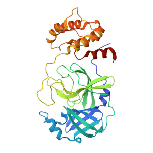
    9DTZ, 9DU2, 9DU3, 9DU4

  • PubMed Abstract: 

    SARS-CoV-2 Mpro is a cysteine protease that acts as a symmetrical dimer and displays positive cooperativity for substrate turnover. A series of potent reversible covalent peptidomimetic aldehydes and nitriles was designed as Mpro inhibitors. To better understand the observed structure activity relationships (SAR), binding potency and mechanism was examined by enzyme activity assay, surface plasmon resonance, X-ray crystallography, matrix-assisted laser desorption electrospray ionization, and nuclear magnetic resonance (NMR). Potent aldehydes bind Mpro cooperativity but bind covalently to only one subunit of the dimer. The analogous nitriles do not bind cooperatively, and the degree of covalent binding observed varied depending on the assay method employed. The NMR studies support that potent inhibition of Mpro by the nitriles does not require covalent binding. The data highlight the caveats in using orthogonal assays to confirm compound mechanism, particularly in cooperative systems.


  • Organizational Affiliation
    • Department of Cancer Biology, Dana-Farber Cancer Institute, Boston, MA 02115, USA.

Macromolecules
Find similar proteins by:  (by identity cutoff)  |  3D Structure
Entity ID: 1
MoleculeChains Sequence LengthOrganismDetailsImage
3C-like proteinase nsp5315Severe acute respiratory syndrome coronavirus 2Mutation(s): 0 
Gene Names: rep1a-1b
EC: 3.4.22.69
UniProt
Find proteins for P0DTD1 (Severe acute respiratory syndrome coronavirus 2)
Explore P0DTD1 
Go to UniProtKB:  P0DTD1
Entity Groups  
Sequence Clusters30% Identity50% Identity70% Identity90% Identity95% Identity100% Identity
UniProt GroupP0DTD1
Sequence Annotations
Expand
  • Reference Sequence
Small Molecules
Ligands 1 Unique
IDChains Name / Formula / InChI Key2D Diagram3D Interactions
A1BCX (Subject of Investigation/LOI)
Query on A1BCX

Download Ideal Coordinates CCD File 
B [auth A]N-[(2S)-3-cyclopropyl-1-({(2R)-1-hydroxy-3-[(3R)-2-oxopyrrolidin-3-yl]propan-2-yl}amino)-1-oxopropan-2-yl]-4-methoxy-1H-indole-2-carboxamide
C23 H30 N4 O5
GUJIFRXGODKXNA-STGLDUPZSA-N
Experimental Data & Validation

Experimental Data

  • Method: X-RAY DIFFRACTION
  • Resolution: 2.07 Å
  • R-Value Free:  0.244 (Depositor), 0.241 (DCC) 
  • R-Value Work:  0.218 (Depositor), 0.214 (DCC) 
  • R-Value Observed: 0.219 (Depositor) 
Space Group: C 1 2 1
Unit Cell:
Length ( Å )Angle ( ˚ )
a = 113.95α = 90
b = 53.64β = 102.614
c = 45.44γ = 90
Software Package:
Software NamePurpose
PHENIXrefinement
autoPROCdata reduction
autoPROCdata scaling
PHENIXphasing

Structure Validation

View Full Validation Report


Ligand Structure Quality Assessment 


Entry History & Funding Information

Deposition Data


Funding OrganizationLocationGrant Number
Not funded--

Revision History  (Full details and data files)

  • Version 1.0: 2025-04-30
    Type: Initial release
  • Version 1.1: 2025-08-06
    Changes: Database references