Phosphorylation-State Modulated Binding of HSP70: Structural Insights and Compensatory Protein Engineering.
Stewart, M., Paththamperuma, C., McCann, C., Cottingim, K., Zhang, H., DelVecchio, R., Peng, I., Fennimore, E., Nix, J.C., Saeed, M.N., George, K., Makaroff, K., Colie, M., Paulakonis, E., Almeida, M.F., Afolayan, A.J., Brown, N.G., Page, R.C., Schisler, J.C.(2025) bioRxiv
- PubMed: 40027613
- DOI: https://doi.org/10.1101/2025.02.17.637997
- Primary Citation of Related Structures:
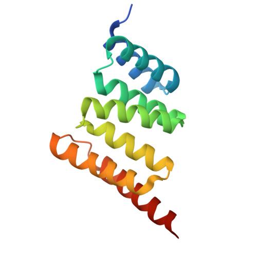
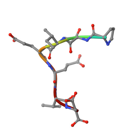
9DYA, 9DYB - PubMed Abstract:
Protein quality control is crucial for cellular homeostasis, involving the heat shock response, the ubiquitin-proteasome system, and the autophagy-lysosome pathway. Central to these systems are the chaperone homologs heat shock protein 70 (HSP70) and heat shock cognate 70 (HSC70), which manage protein folding and degradation. This study investigated the impact of the C-terminal phosphorylation of HSP70 on its interaction with the co-chaperone CHIP (C-terminus of HSC70 interacting protein), an E3 ligase that ubiquitinates protein substrates for degradation. Using both cell-free and cell-based approaches, including X-ray crystallography, biolayer interferometry, and live cell biocomplementation assays, we demonstrate that phosphorylation at HSP70 T636 reduces CHIP's binding affinity, shifting the preference toward other co-chaperones like HOP. Structural analysis reveals that phosphorylation disrupts key hydrogen bonds, altering binding dynamics. We engineered a CHIP variant (CHIP-G132N) to restore binding affinity to phosphorylated HSP70. While CHIP-G132N effectively restored binding without additional functional domains, its effectiveness was diminished in full-length phosphomimetic constructs in cell-free and in-cell assays, suggesting that additional interactions may influence binding. Functional assays indicate that phosphorylation of HSP70 affects its stability and degradation, with implications for diseases such as cancer and neurodegeneration. Our findings highlight the complexity of chaperone-co-chaperone interactions and underscore the importance of post-translational modifications in regulating protein quality control mechanisms. By elucidating the molecular details of HSP70 and CHIP interactions, our study provides a foundation for developing therapeutic interventions for diseases characterized by proteostasis imbalance.
Organizational Affiliation:
The McAllister Heart Institute, The University of North Carolina at Chapel Hill, Chapel Hill, NC 27599, USA.