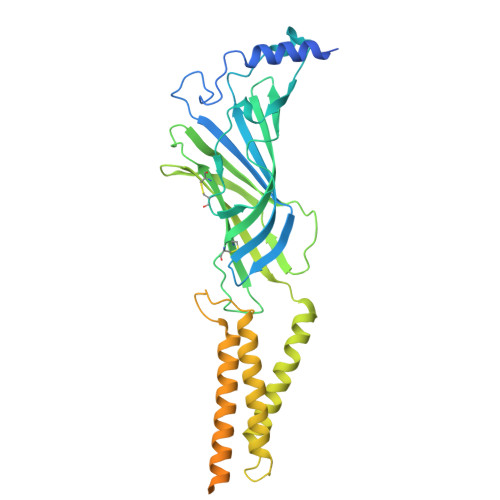
9E6C | pdb_00009e6c

Octopus sensory receptor CRT1 in complex with H3C


Experimental Data Snapshot

  • Method: ELECTRON MICROSCOPY
  • Resolution: 3.04 Å
  • Aggregation State: PARTICLE 
  • Reconstruction Method: SINGLE PARTICLE 

wwPDB Validation   3D Report Full Report


This is version 1.1 of the entry. See complete history


Literature

Environmental microbiomes drive chemotactile sensation in octopus.

Sepela, R.J.Jiang, H.Shin, Y.H.Hautala, T.L.Clardy, J.Hibbs, R.E.Bellono, N.W.

(2025) Cell 188: 4849

  • DOI: https://doi.org/10.1016/j.cell.2025.05.033
  • Primary Citation of Related Structures:  
    9E6B, 9E6C, 9E6D

  • PubMed Abstract: 

    Microbial communities coat nearly every surface in the environment and have co-existed with animals throughout evolution. Whether animals exploit omnipresent microbial cues to navigate their surroundings is not well understood. Octopuses use "taste-by-touch" chemotactile receptors (CRs) to explore the seafloor, but how they distinguish meaningful surfaces from the rocks and crevices they encounter is unknown. Here, we report that secreted signals from microbiomes of ecologically relevant surfaces activate CRs to guide octopus behavior. Distinct molecules isolated from individual bacterial strains located on prey or eggs bind single CRs in subtly different structural conformations to elicit specific mechanisms of receptor activation, ion permeation and signal transduction, and maternal care and predation behavior. Thus, microbiomes on ecological surfaces act at the level of primary sensory receptors to inform behavior. Our study demonstrates that uncovering interkingdom interactions is essential to understanding how animal sensory systems evolved in a microbe-rich world.


  • Organizational Affiliation
    • Department of Molecular and Cellular Biology, Harvard University, Cambridge, MA, USA.

Macromolecules
Find similar proteins by:  (by identity cutoff)  |  3D Structure
Entity ID: 1
MoleculeChains Sequence LengthOrganismDetailsImage
Chemotactile receptor CRT1
A, B, C, D, E
410Octopus bimaculoidesMutation(s): 0 
Gene Names: OCBIM_22006518mg
UniProt
Find proteins for A0A0L8FVQ9 (Octopus bimaculoides)
Explore A0A0L8FVQ9 
Go to UniProtKB:  A0A0L8FVQ9
Entity Groups  
Sequence Clusters30% Identity50% Identity70% Identity90% Identity95% Identity100% Identity
UniProt GroupA0A0L8FVQ9
Glycosylation
Glycosylation Sites: 4
Sequence Annotations
Expand
  • Reference Sequence
Small Molecules
Ligands 2 Unique
IDChains Name / Formula / InChI Key2D Diagram3D Interactions
A1BE4 (Subject of Investigation/LOI)
Query on A1BE4

Download Ideal Coordinates CCD File 
DA [auth E],
J [auth A],
O [auth B],
T [auth C],
Y [auth D]
1-methyl-9H-pyrido[3,4-b]indole-3-carboxylic acid
C13 H10 N2 O2
MFEZJNMQTQMDRQ-UHFFFAOYSA-N
NAG
Query on NAG

Download Ideal Coordinates CCD File 
AA [auth E]
BA [auth E]
CA [auth E]
F [auth A]
G [auth A]
AA [auth E],
BA [auth E],
CA [auth E],
F [auth A],
G [auth A],
H [auth A],
I [auth A],
K [auth B],
L [auth B],
M [auth B],
N [auth B],
P [auth C],
Q [auth C],
R [auth C],
S [auth C],
U [auth D],
V [auth D],
W [auth D],
X [auth D],
Z [auth E]
2-acetamido-2-deoxy-beta-D-glucopyranose
C8 H15 N O6
OVRNDRQMDRJTHS-FMDGEEDCSA-N
Experimental Data & Validation

Experimental Data

  • Method: ELECTRON MICROSCOPY
  • Resolution: 3.04 Å
  • Aggregation State: PARTICLE 
  • Reconstruction Method: SINGLE PARTICLE 
EM Software:
TaskSoftware PackageVersion
MODEL REFINEMENTPHENIX

Structure Validation

View Full Validation Report


Entry History & Funding Information

Deposition Data


Funding OrganizationLocationGrant Number
National Institutes of Health/National Institute of Neurological Disorders and Stroke (NIH/NINDS)United States--

Revision History  (Full details and data files)

  • Version 1.0: 2025-07-09
    Type: Initial release
  • Version 1.1: 2025-09-17
    Changes: Data collection, Database references