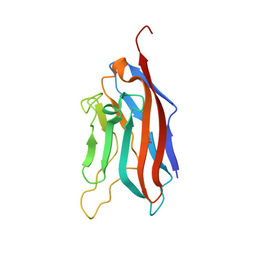
9E6Y | pdb_00009e6y

Structure of CD112 (Nectin-2) domain 1 bound to CD112R (PVRIG)


Experimental Data Snapshot

  • Method: X-RAY DIFFRACTION
  • Resolution: 2.20 Å
  • R-Value Free: 
    0.245 (Depositor), 0.246 (DCC) 
  • R-Value Work: 
    0.208 (Depositor), 0.207 (DCC) 
  • R-Value Observed: 
    0.210 (Depositor) 

Starting Models: experimental, in silico
View more details

wwPDB Validation   3D Report Full Report


Ligand Structure Quality Assessment 


This is version 1.1 of the entry. See complete history


Literature

Structure-guided engineering of CD112 receptor variants for optimized immunotherapy.

Singh, S.Julia, E.Kalita, P.Mason, C.Ming, Q.Lee-Sam, A.Gordon, S.Buitrago, M.E.Leung, D.W.Hwu, P.Luca, V.C.

(2025) Mol Ther 33: 3590-3604

  • DOI: https://doi.org/10.1016/j.ymthe.2025.04.032
  • Primary Citation of Related Structures:  
    9E6Y

  • PubMed Abstract: 

    The immune checkpoint protein, CD112 receptor (CD112R, also known as PVRIG), suppresses T and NK cell activation upon binding to tumor-expressed CD112 (Nectin-2) ligands. Here, we determine the structure of the CD112-CD112R complex and use it to guide the engineering of multiple CD112-targeting immunotherapy candidates. The 2.2 Å-resolution crystal structure reveals an antiparallel, lock-and-key binding mode in which CD112R disrupts CD112 homodimerization. Structural analysis informed directed evolution campaigns focused on remodeling the CD112-CD112R interface, resulting in the isolation of CD112R mutants with greatly increased expression and CD112-binding affinity. The highest-affinity variant, CD112R IVE , potently inhibits CD112-CD112R interactions when utilized as a soluble CD112 trap. Furthermore, incorporating CD112R variants into chimeric antigen receptors (CARs) and T cell engagers (TCEs) leads to more robust T cell activation and killing of CD112 + triple-negative breast cancer (TNBC) cells compared to wild-type CD112R. This strategy demonstrates how structural insights can be leveraged to efficiently generate panels of "affinity-tuned" biologics for immunotherapy.


  • Organizational Affiliation
    • Department of Immunology, H. Lee Moffitt Cancer Center and Research Institute, Tampa, FL, 33612, USA; Cancer Biology Ph.D. Program, University of South Florida, Tampa, FL 33612, USA.

Macromolecules
Find similar proteins by:  (by identity cutoff)  |  3D Structure
Entity ID: 1
MoleculeChains Sequence LengthOrganismDetailsImage
Nectin-2A [auth D]132Homo sapiensMutation(s): 0 
Gene Names: NECTIN2HVEBPRR2PVRL2
UniProt & NIH Common Fund Data Resources
Find proteins for Q92692 (Homo sapiens)
Explore Q92692 
Go to UniProtKB:  Q92692
PHAROS:  Q92692
GTEx:  ENSG00000130202 
Entity Groups  
Sequence Clusters30% Identity50% Identity70% Identity90% Identity95% Identity100% Identity
UniProt GroupQ92692
Sequence Annotations
Expand
  • Reference Sequence
Find similar proteins by:  (by identity cutoff)  |  3D Structure
Entity ID: 2
MoleculeChains Sequence LengthOrganismDetailsImage
Transmembrane protein PVRIGB [auth A]133Homo sapiensMutation(s): 0 
Gene Names: PVRIGC7orf15
UniProt & NIH Common Fund Data Resources
Find proteins for Q6DKI7 (Homo sapiens)
Explore Q6DKI7 
Go to UniProtKB:  Q6DKI7
GTEx:  ENSG00000213413 
Entity Groups  
Sequence Clusters30% Identity50% Identity70% Identity90% Identity95% Identity100% Identity
UniProt GroupQ6DKI7
Glycosylation
Glycosylation Sites: 1Go to GlyGen: Q6DKI7-1
Sequence Annotations
Expand
  • Reference Sequence
Small Molecules
Ligands 2 Unique
IDChains Name / Formula / InChI Key2D Diagram3D Interactions
NAG (Subject of Investigation/LOI)
Query on NAG

Download Ideal Coordinates CCD File 
I [auth A]2-acetamido-2-deoxy-beta-D-glucopyranose
C8 H15 N O6
OVRNDRQMDRJTHS-FMDGEEDCSA-N
EDO (Subject of Investigation/LOI)
Query on EDO

Download Ideal Coordinates CCD File 
C [auth D]
D
E [auth D]
F [auth D]
G [auth D]
C [auth D],
D,
E [auth D],
F [auth D],
G [auth D],
H [auth D],
J [auth A],
K [auth A],
L [auth A],
M [auth A],
N [auth A]
1,2-ETHANEDIOL
C2 H6 O2
LYCAIKOWRPUZTN-UHFFFAOYSA-N
Experimental Data & Validation

Experimental Data

  • Method: X-RAY DIFFRACTION
  • Resolution: 2.20 Å
  • R-Value Free:  0.245 (Depositor), 0.246 (DCC) 
  • R-Value Work:  0.208 (Depositor), 0.207 (DCC) 
  • R-Value Observed: 0.210 (Depositor) 
Space Group: P 43 21 2
Unit Cell:
Length ( Å )Angle ( ˚ )
a = 79.65α = 90
b = 79.65β = 90
c = 81.38γ = 90
Software Package:
Software NamePurpose
PHENIXrefinement
PHENIXrefinement
XDSdata reduction
XDSdata scaling
PHENIXphasing

Structure Validation

View Full Validation Report


Ligand Structure Quality Assessment 


Entry History & Funding Information

Deposition Data


Funding OrganizationLocationGrant Number
National Institutes of Health/National Institute of General Medical Sciences (NIH/NIGMS)United StatesGM133482-01

Revision History  (Full details and data files)

  • Version 1.0: 2025-05-07
    Type: Initial release
  • Version 1.1: 2025-08-20
    Changes: Database references