9EMQ | pdb_00009emq

Crystal Structure of DC-SIGN in complex with AL86


Experimental Data Snapshot

  • Method: X-RAY DIFFRACTION
  • Resolution: 1.80 Å
  • R-Value Free: 
    0.197 (Depositor), 0.197 (DCC) 
  • R-Value Work: 
    0.167 (Depositor), 0.167 (DCC) 
  • R-Value Observed: 
    0.169 (Depositor) 

Starting Model: experimental
View more details

wwPDB Validation   3D Report Full Report


Ligand Structure Quality Assessment 


This is version 1.0 of the entry. See complete history


Literature

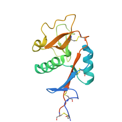
Thermodynamics-Guided Design Reveals a Cooperative Hydrogen Bond in DC-SIGN-targeted Glycomimetics.

Nemli, D.D.Jiang, X.Jakob, R.P.Gloder, L.M.Schwardt, O.Rabbani, S.Maier, T.Ernst, B.Cramer, J.

(2024) J Med Chem 67: 13813-13828

  • DOI: https://doi.org/10.1021/acs.jmedchem.4c00623
  • Primary Citation of Related Structures:  
    9EMQ, 9EMR, 9EMS

  • PubMed Abstract: 

    Due to the shallow and hydrophilic binding sites of carbohydrate-binding proteins, the design of glycomimetics is often complicated by high desolvation costs as well as competition with solvent. Therefore, a careful optimization of interaction vectors and ligand properties is required in the design and optimization of glycomimetics. Here, we employ thermodynamics-guided design to optimize mannose-based glycomimetics targeting the human C-type lectin receptor dendritic cell-specific intercellular adhesion molecule 3 grabbing nonintegrin (DC-SIGN), a pathogenic host factor in viral infections. By exploring ligand rigidification and hydrogen bond engineering, a monovalent glycomimetic with an unprecedented affinity for DC-SIGN in the low μM range was discovered. A matched molecular pair analysis based on microcalorimetric data revealed a stereospecific hydrogen bond interaction with Glu358/Ser360 as the origin of this cooperative and enthalpically dominated interaction. This detailed insight into the binding mechanism paves the way for an improvement of monovalent glycomimetics targeting DC-SIGN.


  • Organizational Affiliation
    • Institute for Pharmaceutical and Medicinal Chemistry, Heinrich-Heine-University Düsseldorf, Universitätsstraße 1, Düsseldorf 40225, Germany.

Macromolecules
Find similar proteins by:  (by identity cutoff)  |  3D Structure
Entity ID: 1
MoleculeChains Sequence LengthOrganismDetailsImage
DC-SIGN, CRD domain159Homo sapiensMutation(s): 0 
Gene Names: CD209CLEC4L
UniProt & NIH Common Fund Data Resources
Find proteins for Q9NNX6 (Homo sapiens)
Explore Q9NNX6 
Go to UniProtKB:  Q9NNX6
PHAROS:  Q9NNX6
GTEx:  ENSG00000090659 
Entity Groups  
Sequence Clusters30% Identity50% Identity70% Identity90% Identity95% Identity100% Identity
UniProt GroupQ9NNX6
Sequence Annotations
Expand
  • Reference Sequence
Experimental Data & Validation

Experimental Data

  • Method: X-RAY DIFFRACTION
  • Resolution: 1.80 Å
  • R-Value Free:  0.197 (Depositor), 0.197 (DCC) 
  • R-Value Work:  0.167 (Depositor), 0.167 (DCC) 
  • R-Value Observed: 0.169 (Depositor) 
Space Group: P 3 2 1
Unit Cell:
Length ( Å )Angle ( ˚ )
a = 59.29α = 90
b = 59.29β = 90
c = 74.295γ = 120
Software Package:
Software NamePurpose
PHENIXrefinement
PDB_EXTRACTdata extraction
XDSdata reduction
Aimlessdata scaling
PHASERphasing

Structure Validation

View Full Validation Report


Ligand Structure Quality Assessment 


Entry History & Funding Information

Deposition Data


Funding OrganizationLocationGrant Number
German Research Foundation (DFG)Germany532758733

Revision History  (Full details and data files)

  • Version 1.0: 2024-11-13
    Type: Initial release