9F2D | pdb_00009f2d

KIR2DL1 bound to RIFIN RBK21


Experimental Data Snapshot

  • Method: X-RAY DIFFRACTION
  • Resolution: 2.89 Å
  • R-Value Free: 
    0.286 (Depositor), 0.257 (DCC) 
  • R-Value Work: 
    0.256 (Depositor), 0.260 (DCC) 
  • R-Value Observed: 
    0.258 (Depositor) 

Starting Model: experimental
View more details

wwPDB Validation   3D Report Full Report


Ligand Structure Quality Assessment 


This is version 1.2 of the entry. See complete history


Literature

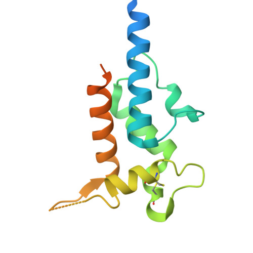
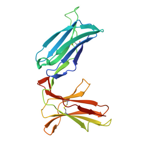
RIFINs displayed on malaria-infected erythrocytes bind KIR2DL1 and KIR2DS1.

Sakoguchi, A.Chamberlain, S.G.Morch, A.M.Widdess, M.Harrison, T.E.Dustin, M.L.Arase, H.Higgins, M.K.Iwanaga, S.

(2025) Nature 

  • DOI: https://doi.org/10.1038/s41586-025-09091-y
  • Primary Citation of Related Structures:  
    9F2D, 9HML

  • PubMed Abstract: 

    Natural killer (NK) cells use inhibitory and activating immune receptors to differentiate between human cells and pathogens. Signalling by these receptors determines whether an NK cell becomes activated and destroys a target cell. In some cases, such as killer immunoglobulin-like receptors, immune receptors are found in pairs, with inhibitory and activating receptors containing nearly identical extracellular ligand-binding domains coupled to different intracellular signalling domains 1 . Previous studies showed that repetitive interspersed family (RIFIN) proteins, displayed on the surfaces of Plasmodium falciparum-infected erythrocytes, can bind to inhibitory immune receptors and dampen NK cell activation 2,3 , reducing parasite killing. However, no pathogen-derived ligand has been identified for any human activating receptor. Here we identified a clade of RIFINs that bind to inhibitory immune receptor KIR2DL1 more strongly than KIR2DL1 binds to the human ligand (MHC class I). This interaction mediates inhibitory signalling and suppresses the activation of KIR2DL1-expressing NK cells. We show that KIR2DL1-binding RIFINs are abundant in field-isolated strains from both Africa and Asia and reveal how the two RIFINs bind to KIR2DL1. The RIFIN binding surface of KIR2DL1 is conserved in the cognate activating immune receptor KIR2DS1. We find that KIR2DL1-binding RIFINs can also bind to KIR2DS1, resulting in the activation of KIR2DS1-expressing NK cells. This study demonstrates that activating killer immunoglobulin-like receptors can recruit NK cells to target a pathogen and reveals a potential role for activating immune receptors in controlling malaria infection.


  • Organizational Affiliation

    Department of Protozoology, Research Institute for Microbial Diseases, The University of Osaka, Suita, Japan.


Macromolecules
Find similar proteins by:  (by identity cutoff)  |  3D Structure
Entity ID: 1
MoleculeChains Sequence LengthOrganismDetailsImage
RIFIN RBK21
A, C, E, G, I
A, C, E, G, I, K, M, O
152Plasmodium falciparumMutation(s): 0 
UniProt
Find proteins for W7F7W4 (Plasmodium falciparum (isolate 7G8))
Explore W7F7W4 
Go to UniProtKB:  W7F7W4
Entity Groups  
Sequence Clusters30% Identity50% Identity70% Identity90% Identity95% Identity100% Identity
UniProt GroupW7F7W4
Glycosylation
Glycosylation Sites: 1
Sequence Annotations
Expand
  • Reference Sequence
Find similar proteins by:  (by identity cutoff)  |  3D Structure
Entity ID: 2
MoleculeChains Sequence LengthOrganismDetailsImage
KIR2DL protein
B, D, F, H, J
B, D, F, H, J, L, N, P
195Homo sapiensMutation(s): 0 
Gene Names: KIR2DL13DL2
UniProt & NIH Common Fund Data Resources
Find proteins for P43626 (Homo sapiens)
Explore P43626 
Go to UniProtKB:  P43626
PHAROS:  P43626
GTEx:  ENSG00000125498 
Entity Groups  
Sequence Clusters30% Identity50% Identity70% Identity90% Identity95% Identity100% Identity
UniProt GroupP43626
Glycosylation
Glycosylation Sites: 1Go to GlyGen: P43626-1
Sequence Annotations
Expand
  • Reference Sequence
Oligosaccharides

Help

Entity ID: 3
MoleculeChains Length2D Diagram Glycosylation3D Interactions
alpha-L-fucopyranose-(1-6)-2-acetamido-2-deoxy-beta-D-glucopyranose
Q, R, S, T, U
2N-Glycosylation
Glycosylation Resources
GlyTouCan:  G86851RC
GlyCosmos:  G86851RC
GlyGen:  G86851RC
Small Molecules
Ligands 1 Unique
IDChains Name / Formula / InChI Key2D Diagram3D Interactions
NAG
Query on NAG

Download Ideal Coordinates CCD File 
AA [auth H]
BA [auth H]
CA [auth I]
DA [auth J]
EA [auth L]
AA [auth H],
BA [auth H],
CA [auth I],
DA [auth J],
EA [auth L],
FA [auth L],
GA [auth N],
HA [auth N],
IA [auth O],
JA [auth P],
V [auth A],
W [auth B],
X [auth D],
Y [auth E],
Z [auth F]
2-acetamido-2-deoxy-beta-D-glucopyranose
C8 H15 N O6
OVRNDRQMDRJTHS-FMDGEEDCSA-N
Experimental Data & Validation

Experimental Data

  • Method: X-RAY DIFFRACTION
  • Resolution: 2.89 Å
  • R-Value Free:  0.286 (Depositor), 0.257 (DCC) 
  • R-Value Work:  0.256 (Depositor), 0.260 (DCC) 
  • R-Value Observed: 0.258 (Depositor) 
Space Group: P 21 21 21
Unit Cell:
Length ( Å )Angle ( ˚ )
a = 93.399α = 90
b = 99.047β = 90
c = 321.13γ = 90
Software Package:
Software NamePurpose
BUSTERrefinement
PDB_EXTRACTdata extraction
DIALSdata reduction
Aimlessdata scaling
PHASERphasing

Structure Validation

View Full Validation Report


Ligand Structure Quality Assessment 


Entry History & Funding Information

Deposition Data


Funding OrganizationLocationGrant Number
Wellcome TrustUnited Kingdom224343/Z/21/Z

Revision History  (Full details and data files)

  • Version 1.0: 2025-04-09
    Type: Initial release
  • Version 1.1: 2025-06-11
    Changes: Database references
  • Version 1.2: 2025-06-25
    Changes: Database references