RING dimerisation drives higher-order organisation of SINA/SIAH E3 ubiquitin ligases.
Coste, F., Mishra, A., Chapuis, C., Mance, L., Pukalo, Z., Bigot, N., Goffinont, S., Gaudon, V., Garnier, N., Talhaoui, I., Castaing, B., Huet, S., Suskiewicz, M.J.(2025) FEBS J 292: 2784-2805
- PubMed: 39910688
- DOI: https://doi.org/10.1111/febs.70000
- Primary Citation of Related Structures:
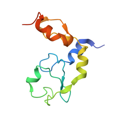
9G0L - PubMed Abstract:
RING-type E3 ubiquitin ligases promote ubiquitylation by stabilising an active complex between a ubiquitin-loaded E2-conjugating enzyme and a protein substrate. To fulfil this function, the E3 ubiquitin-protein ligase SIAH1 and other SINA/SIAH subfamily RING-type E3 ligases employ an N-terminal catalytic RING domain and a C-terminal substrate-binding domain (SBD), separated by two zinc fingers. Here, we present the first crystal structure of the RING domain of human SIAH1, together with an adjacent zinc finger, revealing a potential RING dimer, which was validated in solution using static light scattering. RING dimerisation contributes to the E3 ligase activity of SIAH1 both in vitro and in cells. Moreover, as the RING domain is the second element, after the SBD, to independently favour homodimerisation within SINA/SIAH E3 ligases, we propose that alternating RING:RING and SBD:SBD interactions organise multiple copies of a SINA/SIAH protein into a higher-order homomultimer. In line with this hypothesis, fluorescently tagged full-length human SIAH1, human SIAH2 and fruit fly SINA show cytoplasmic clusters in human cells, whereas their distribution becomes more diffuse when RING dimerisation is disabled. The wild-type (WT) form of SIAH1, but not its RING dimerisation mutant, colocalises with aggregated synphilin-1A under proteasomal inhibition, suggesting that SIAH1 multimerisation might contribute to its reported preference for aggregated or multimeric substrates.
- Centre de Biophysique Moléculaire (CBM), UPR 4301, CNRS, Orléans, France.
Organizational Affiliation: