9G6T | pdb_00009g6t

p53-Y220C Core Domain Covalently Bound to 5-Chloro-6-methylpyrazine-2-carbonitrile Soaked at 5 mM


Experimental Data Snapshot

  • Method: X-RAY DIFFRACTION
  • Resolution: 1.56 Å
  • R-Value Free: 
    0.199 (Depositor), 0.210 (DCC) 
  • R-Value Work: 
    0.150 (Depositor), 0.169 (DCC) 
  • R-Value Observed: 
    0.151 (Depositor) 

Starting Model: experimental
View more details

wwPDB Validation   3D Report Full Report


Ligand Structure Quality Assessment 


This is version 1.1 of the entry. See complete history


Literature

S N Ar Reactive Pyrazine Derivatives as p53-Y220C Cleft Binders with Diverse Binding Modes.

Klett, T.Stahlecker, J.Schwer, M.Jaag, S.J.Masberg, B.Knappe, C.Lammerhofer, M.Stehle, T.Boeckler, F.M.

(2025) Drug Des Devel Ther 19: 4727-4753

  • DOI: https://doi.org/10.2147/DDDT.S513792
  • Primary Citation of Related Structures:  
    9G5H, 9G6T, 9G6U

  • PubMed Abstract: 

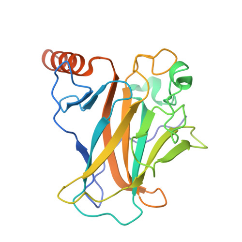
    The tumor suppressor p53 is most commonly mutated in human cancer. The structural mutant in the β-sandwich of the protein, p53-Y220C, is the ninth most common p53 mutant. The p53-Y220C mutant has a solvent-accessible hydrophobic pocket, leading to thermal destabilization of the protein. Screening of our covalent fragment library (CovLib) revealed the highly reactive pyrazine derivatives SN006 and SN007, which arylate among other cysteines in p53, the mutation-generated Cys220. Herein, comprehensive structure-activity relationship (SAR) studies of these intrinsically reactive CovLib hits were performed, aiming to identify improved stabilizers for p53-Y220C, with a more balanced reactivity profile, diverse binding modes and a better potential for chemical optimization. The compounds were screened for enhanced stabilization of p53 wild type and its mutants using differential scanning fluorimetry (DSF). To confirm covalent modification, intact mass spectrometry was performed. Thiol reactivity profiles were determined using a standardized Glutathione-modifying (GSH) assay. The binding modes of the identified hits and covalent modification of Cys220 were elucidated by X-ray crystallography. Moreover, the influence of the hits on the DNA-binding affinity of full-length p53 was investigated employing a fluorescence polarization assay (FPA). The promising pyrazine derivatives SN006/7-3, SN006/7-8, and SN006/7-9 were identified, occupying different subsites of the Y220C binding pocket. The compound SN006/7-8 substantially stabilized the thermosensitive cancer mutant Y220C by up to 5.0 °C, representing a strong enhancement over SN006 (1.8 °C) and SN007 (2.0 °C).


  • Organizational Affiliation
    • Laboratory for Molecular Design & Pharmaceutical Biophysics, Institute of Pharmaceutical Sciences, Department of Pharmacy and Biochemistry, Eberhard Karls Universität Tübingen, Tübingen, 72076, Germany.

Macromolecules
Find similar proteins by:  (by identity cutoff)  |  3D Structure
Entity ID: 1
MoleculeChains Sequence LengthOrganismDetailsImage
Cellular tumor antigen p53
A, B
219Homo sapiensMutation(s): 5 
Gene Names: TP53P53
UniProt & NIH Common Fund Data Resources
Find proteins for P04637 (Homo sapiens)
Explore P04637 
Go to UniProtKB:  P04637
PHAROS:  P04637
GTEx:  ENSG00000141510 
Entity Groups  
Sequence Clusters30% Identity50% Identity70% Identity90% Identity95% Identity100% Identity
UniProt GroupP04637
Sequence Annotations
Expand
  • Reference Sequence
Small Molecules
Ligands 6 Unique
IDChains Name / Formula / InChI Key2D Diagram3D Interactions
EPE
Query on EPE

Download Ideal Coordinates CCD File 
F [auth A],
M [auth A],
T [auth B]
4-(2-HYDROXYETHYL)-1-PIPERAZINE ETHANESULFONIC ACID
C8 H18 N2 O4 S
JKMHFZQWWAIEOD-UHFFFAOYSA-N
A1II4 (Subject of Investigation/LOI)
Query on A1II4

Download Ideal Coordinates CCD File 
C [auth A],
D [auth A],
O [auth B],
P [auth B],
R [auth B]
5-chloranyl-6-methyl-pyrazine-2-carbonitrile
C6 H4 Cl N3
ADCSDJITDCCGAR-UHFFFAOYSA-N
PEG
Query on PEG

Download Ideal Coordinates CCD File 
H [auth A],
Q [auth B],
V [auth B]
DI(HYDROXYETHYL)ETHER
C4 H10 O3
MTHSVFCYNBDYFN-UHFFFAOYSA-N
GOL
Query on GOL

Download Ideal Coordinates CCD File 
J [auth A],
U [auth B],
W [auth B]
GLYCEROL
C3 H8 O3
PEDCQBHIVMGVHV-UHFFFAOYSA-N
ZN
Query on ZN

Download Ideal Coordinates CCD File 
N [auth A],
X [auth B]
ZINC ION
Zn
PTFCDOFLOPIGGS-UHFFFAOYSA-N
EDO
Query on EDO

Download Ideal Coordinates CCD File 
E [auth A]
G [auth A]
I [auth A]
K [auth A]
L [auth A]
E [auth A],
G [auth A],
I [auth A],
K [auth A],
L [auth A],
S [auth B]
1,2-ETHANEDIOL
C2 H6 O2
LYCAIKOWRPUZTN-UHFFFAOYSA-N
Experimental Data & Validation

Experimental Data

  • Method: X-RAY DIFFRACTION
  • Resolution: 1.56 Å
  • R-Value Free:  0.199 (Depositor), 0.210 (DCC) 
  • R-Value Work:  0.150 (Depositor), 0.169 (DCC) 
  • R-Value Observed: 0.151 (Depositor) 
Space Group: P 21 21 21
Unit Cell:
Length ( Å )Angle ( ˚ )
a = 65.191α = 90
b = 71.145β = 90
c = 105.73γ = 90
Software Package:
Software NamePurpose
PHENIXrefinement
XDSdata reduction
XDSdata scaling
PHASERphasing

Structure Validation

View Full Validation Report


Ligand Structure Quality Assessment 


Entry History & Funding Information

Deposition Data


Funding OrganizationLocationGrant Number
Not funded--

Revision History  (Full details and data files)

  • Version 1.0: 2025-06-11
    Type: Initial release
  • Version 1.1: 2025-07-16
    Changes: Database references