9GH5 | pdb_00009gh5

Complex of Fusobacterium nucleatum CbpF with human CEACAM1


Experimental Data Snapshot

  • Method: ELECTRON MICROSCOPY
  • Resolution: 2.70 Å
  • Aggregation State: PARTICLE 
  • Reconstruction Method: SINGLE PARTICLE 

wwPDB Validation   3D Report Full Report


This is version 1.2 of the entry. See complete history


Literature

Structural basis for immune cell binding of Fusobacterium nucleatum via the trimeric autotransporter adhesin CbpF.

Marongiu, G.L.Fink, U.Schopf, F.Oder, A.von Kries, J.P.Roderer, D.

(2025) Proc Natl Acad Sci U S A 122: e2418155122-e2418155122

  • DOI: https://doi.org/10.1073/pnas.2418155122
  • Primary Citation of Related Structures:  
    9GH4, 9GH5, 9GH6

  • PubMed Abstract: 

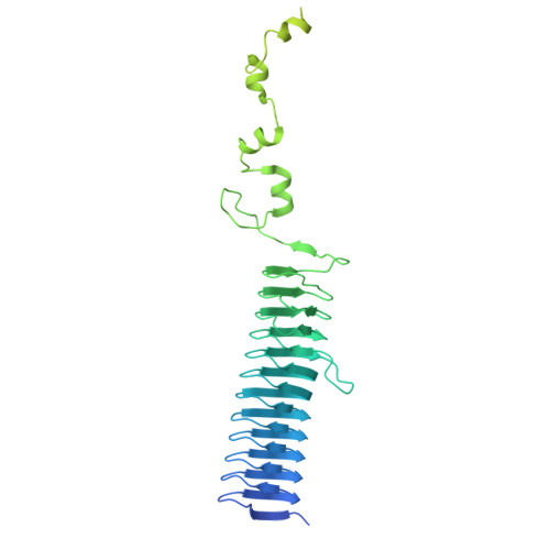
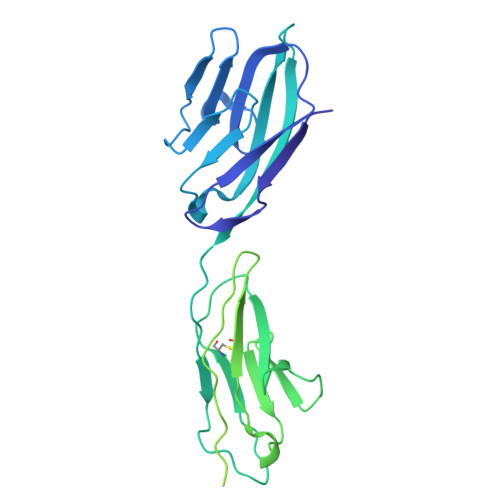
    Fusobacterium nucleatum (Fn), a commensal in the human oral cavity, is overrepresented in the colon microbiota of colorectal cancer (CRC) patients and is linked to tumor chemoresistance, metastasis, and a poor therapeutic prognosis. Fn produces numerous adhesins that mediate tumor colonization and downregulation of the host's antitumor immune response. One of these, the trimeric autotransporter adhesin (TAA) CEACAM binding protein of Fusobacterium (CbpF), targets CEACAM1 on T-cells and has been associated with immune evasion of Fn-colonized tumors. Whereas the role of CEACAM1 in homophilic and heterophilic cell interactions and immune evasion is well described, the mechanistic details of its interaction with fusobacterial CbpF remain unknown due to the lack of a high-resolution structure of the adhesin-receptor complex. Here, we present two structures of CbpF alone and in complex with CEACAM1, obtained by cryogenic electron microscopy and single particle analysis. They reveal that CbpF forms a stable homotrimeric complex whose N-terminal part of the extracellular domain comprises a 64 Å long β roll domain with a unique lateral loop extension. CEACAM1 binds to this loop with high affinity via its N-terminal IgV-like domain with a nanomolar dissociation constant as determined by surface plasmon resonance. This study provides a comprehensive structural description of a fusobacterial TAA, illustrates a yet undescribed CEACAM1 binding mode, and paves the way for rational drug design targeting Fn in CRC.


  • Organizational Affiliation
    • Leibniz-Forschungsinstitut fur Molekulare Pharmakologie, Berlin 13125, Germany.

Macromolecules
Find similar proteins by:  (by identity cutoff)  |  3D Structure
Entity ID: 1
MoleculeChains Sequence LengthOrganismDetailsImage
Cell surface protein
A, B, C
489Fusobacterium nucleatumMutation(s): 0 
Gene Names: FN1499
UniProt
Find proteins for Q8RIS0 (Fusobacterium nucleatum subsp. nucleatum (strain ATCC 25586 / DSM 15643 / BCRC 10681 / CIP 101130 / JCM 8532 / KCTC 2640 / LMG 13131 / VPI 4355))
Explore Q8RIS0 
Go to UniProtKB:  Q8RIS0
Entity Groups  
Sequence Clusters30% Identity50% Identity70% Identity90% Identity95% Identity100% Identity
UniProt GroupQ8RIS0
Sequence Annotations
Expand
  • Reference Sequence
Find similar proteins by:  (by identity cutoff)  |  3D Structure
Entity ID: 2
MoleculeChains Sequence LengthOrganismDetailsImage
Carcinoembryonic antigen-related cell adhesion molecule 1
D, E, F
400Homo sapiensMutation(s): 0 
Gene Names: CEACAM1BGPBGP1
UniProt & NIH Common Fund Data Resources
Find proteins for P13688 (Homo sapiens)
Explore P13688 
Go to UniProtKB:  P13688
PHAROS:  P13688
GTEx:  ENSG00000079385 
Entity Groups  
Sequence Clusters30% Identity50% Identity70% Identity90% Identity95% Identity100% Identity
UniProt GroupP13688
Glycosylation
Glycosylation Sites: 6Go to GlyGen: P13688-1
Sequence Annotations
Expand
  • Reference Sequence
Small Molecules
Ligands 1 Unique
IDChains Name / Formula / InChI Key2D Diagram3D Interactions
NAG
Query on NAG

Download Ideal Coordinates CCD File 
G [auth D]
H [auth D]
I [auth D]
J [auth D]
K [auth D]
G [auth D],
H [auth D],
I [auth D],
J [auth D],
K [auth D],
L [auth D],
M [auth E],
N [auth E],
O [auth E],
P [auth E],
Q [auth E],
R [auth E],
S [auth F],
T [auth F],
U [auth F],
V [auth F],
W [auth F],
X [auth F]
2-acetamido-2-deoxy-beta-D-glucopyranose
C8 H15 N O6
OVRNDRQMDRJTHS-FMDGEEDCSA-N
Experimental Data & Validation

Experimental Data

  • Method: ELECTRON MICROSCOPY
  • Resolution: 2.70 Å
  • Aggregation State: PARTICLE 
  • Reconstruction Method: SINGLE PARTICLE 
EM Software:
TaskSoftware PackageVersion
RECONSTRUCTIONcryoSPARC4.4.0
MODEL REFINEMENTPHENIX1.21-5207

Structure Validation

View Full Validation Report


Entry History & Funding Information

Deposition Data


Funding OrganizationLocationGrant Number
Leibniz AssociationGermany--

Revision History  (Full details and data files)

  • Version 1.0: 2025-04-02
    Type: Initial release
  • Version 1.1: 2025-04-16
    Changes: Data collection, Database references
  • Version 1.2: 2025-04-23
    Changes: Data collection, Database references