9GV8 | pdb_00009gv8

N-Acyl-D-amino-acid deacylase (D-acylase) from Klebsiella pneumoniae in the absence of glycerol


Experimental Data Snapshot

  • Method: X-RAY DIFFRACTION
  • Resolution: 2.60 Å
  • R-Value Free: 
    0.269 (Depositor), 0.266 (DCC) 
  • R-Value Work: 
    0.226 (Depositor), 0.224 (DCC) 
  • R-Value Observed: 
    0.228 (Depositor) 

Starting Model: experimental
View more details

wwPDB Validation   3D Report Full Report


Ligand Structure Quality Assessment 


This is version 1.0 of the entry. See complete history


Literature

Revisiting D-Acylases for D-Amino Acid Production.

Martinez-Rodriguez, S.Gavira, J.A.

(2025) Microb Biotechnol 18: e70179-e70179

  • DOI: https://doi.org/10.1111/1751-7915.70179
  • Primary Citation of Related Structures:  
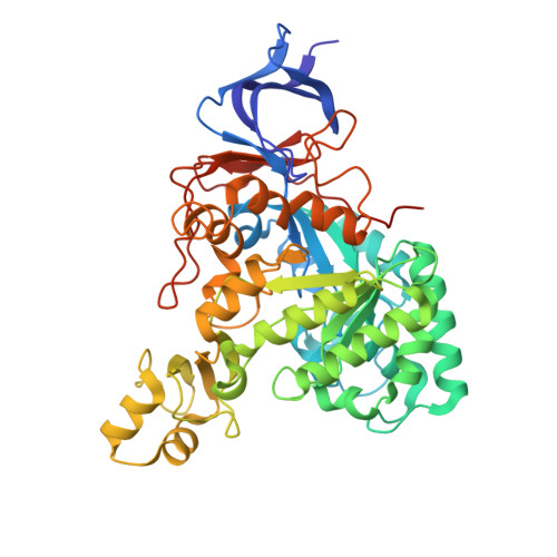
    9G5M, 9GV8

  • PubMed Abstract: 

    N-Acyl-D-amino acid deacylases (EC 3.5.1.81, also known as D-acylases) have been studied for decades for their utility in the kinetic resolution of N-acetyl-D,L-amino acids (NAAs) due to a marked stereospecificity. In conjunction with an N-succinyl-amino acid racemase (NSAR), they impulse the dynamic kinetic resolution (DKR) of different NAAs until the corresponding enantiomerically pure D-amino acids. Besides the clear interest in this enzyme cascade, the application of D-acylase/NSAR tandems has been only briefly described outside the industrial field. In this work, we revisit D-acylases for the DKR of NAAs, reporting the characterisation of two new recombinant D-acylases belonging to Bordetella petrii and Klebsiella pneumoniae. The enzymes were successfully coupled with the recombinant NSAR from Geobacillus stearothermophilus for the biosynthesis of D-methionine or D-aminobutyric acid. We also carried out the structural characterisation of the D-acylase from Klebsiella pneumoniae (KleDacyl), providing the second experimental 3-D structure of a member of this family of enzymes. The structural model shows a highly dynamic character of this amidohydrolase superfamily member, supplying a snapshot of an open conformation of the enzyme most likely preceding substrate entrance into the catalytic cleft. Our results confirm for the first time the importance of an α/β mobile domain in the substrate specificity of D-acylases (region 282-341 in KleDacyl), opening up new strategies for structural-based protein engineering strategies.


  • Organizational Affiliation
    • Department of Biochemistry and Molecular Biology III and Immunology, University of Granada, Granada, Spain.

Macromolecules
Find similar proteins by:  (by identity cutoff)  |  3D Structure
Entity ID: 1
MoleculeChains Sequence LengthOrganismDetailsImage
Amidohydrolase family protein485Klebsiella pneumoniae subsp. pneumoniae Kp13Mutation(s): 0 
Gene Names: 
EC: 3.5.1 (PDB Primary Data), 3.5.1.81 (PDB Primary Data)
UniProt
Find proteins for W8VI54 (Klebsiella pneumoniae 30684/NJST258_2)
Explore W8VI54 
Go to UniProtKB:  W8VI54
Entity Groups  
Sequence Clusters30% Identity50% Identity70% Identity90% Identity95% Identity100% Identity
UniProt GroupW8VI54
Sequence Annotations
Expand
  • Reference Sequence
Small Molecules
Ligands 3 Unique
IDChains Name / Formula / InChI Key2D Diagram3D Interactions
SO4 (Subject of Investigation/LOI)
Query on SO4

Download Ideal Coordinates CCD File 
F [auth A],
G [auth A],
H [auth A],
I [auth A]
SULFATE ION
O4 S
QAOWNCQODCNURD-UHFFFAOYSA-L
EDO
Query on EDO

Download Ideal Coordinates CCD File 
B [auth A],
C [auth A],
D [auth A],
E [auth A]
1,2-ETHANEDIOL
C2 H6 O2
LYCAIKOWRPUZTN-UHFFFAOYSA-N
NI (Subject of Investigation/LOI)
Query on NI

Download Ideal Coordinates CCD File 
J [auth A],
K [auth A]
NICKEL (II) ION
Ni
VEQPNABPJHWNSG-UHFFFAOYSA-N
Experimental Data & Validation

Experimental Data

  • Method: X-RAY DIFFRACTION
  • Resolution: 2.60 Å
  • R-Value Free:  0.269 (Depositor), 0.266 (DCC) 
  • R-Value Work:  0.226 (Depositor), 0.224 (DCC) 
  • R-Value Observed: 0.228 (Depositor) 
Space Group: P 32 2 1
Unit Cell:
Length ( Å )Angle ( ˚ )
a = 101.422α = 90
b = 101.422β = 90
c = 124.94γ = 120
Software Package:
Software NamePurpose
PHENIXrefinement
Aimlessdata scaling
xia2data reduction
MOLREPphasing

Structure Validation

View Full Validation Report


Ligand Structure Quality Assessment 


Entry History & Funding Information

Deposition Data


Funding OrganizationLocationGrant Number
Ministerio de Ciencia e Innovacion (MCIN)SpainPID2020-116261GB-I00

Revision History  (Full details and data files)

  • Version 1.0: 2025-06-18
    Type: Initial release