


Crystal structure of human geranylgeranyl diphosphate synthase mutant R235C
Yehia, R., Kadek, A., Portasikova, J.M., Man, P., Giladi, M., Haitin, Y.To be published.
Experimental Data Snapshot
Starting Model: experimental
View more details
Entity ID: 1 | |||||
|---|---|---|---|---|---|
| Molecule | Chains | Sequence Length | Organism | Details | Image |
| Geranylgeranyl pyrophosphate synthase | 307 | Homo sapiens | Mutation(s): 2 Gene Names: GGPS1 EC: 2.5.1 (PDB Primary Data), 2.5.1.1 (PDB Primary Data), 2.5.1.29 (PDB Primary Data), 2.5.1.10 (PDB Primary Data) | 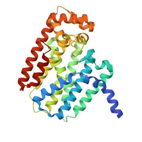 | |
UniProt & NIH Common Fund Data Resources | |||||
Find proteins for O95749 (Homo sapiens) Explore O95749 Go to UniProtKB: O95749 | |||||
PHAROS: O95749 GTEx: ENSG00000152904 | |||||
Entity Groups | |||||
| Sequence Clusters | 30% Identity50% Identity70% Identity90% Identity95% Identity100% Identity | ||||
| UniProt Group | O95749 | ||||
Sequence AnnotationsExpand | |||||
| |||||
| Ligands 2 Unique | |||||
|---|---|---|---|---|---|
| ID | Chains | Name / Formula / InChI Key | 2D Diagram | 3D Interactions | |
| GRG (Subject of Investigation/LOI) Query on GRG | G [auth A] J [auth B] M [auth C] P [auth D] S [auth E] | GERANYLGERANYL DIPHOSPHATE C20 H36 O7 P2 OINNEUNVOZHBOX-QIRCYJPOSA-N |  | ||
| MG (Subject of Investigation/LOI) Query on MG | H [auth A] I [auth A] K [auth B] L [auth B] N [auth C] | MAGNESIUM ION Mg JLVVSXFLKOJNIY-UHFFFAOYSA-N |  | ||
| Length ( Å ) | Angle ( ˚ ) |
|---|---|
| a = 102.623 | α = 90 |
| b = 69.081 | β = 97.39 |
| c = 154.987 | γ = 90 |
| Software Name | Purpose |
|---|---|
| PHENIX | refinement |
| XDS | data reduction |
| XDS | data scaling |
| PHENIX | phasing |
| Funding Organization | Location | Grant Number |
|---|---|---|
| Israel Science Foundation | Israel | 1653/21 |
| Israel Science Foundation | Israel | 1721/16 |
| Other private | Israel | ICRF 01214 |
| Other private | Israel | ICRF 19202 |
| Other private | Israel | Israel Cancer Association 20230029 |
| Other private | Israel | Kahn Foundation's Orion project |
| Other private | Israel | Karl and Leonora Fingerhut Fund for Cancer Research |
| Other private | Israel | Recanati Foundation |


