9IPR | pdb_00009ipr

Crystal structure of CTB10-M1


Experimental Data Snapshot

  • Method: X-RAY DIFFRACTION
  • Resolution: 1.94 Å
  • R-Value Free: 
    0.226 (Depositor), 0.227 (DCC) 
  • R-Value Work: 
    0.191 (Depositor), 0.194 (DCC) 
  • R-Value Observed: 
    0.193 (Depositor) 

Starting Model: experimental
View more details

wwPDB Validation   3D Report Full Report


This is version 1.0 of the entry. See complete history


Literature

Light-Driven Deracemization by a Designed Photoenzyme.

Li, M.Zhang, Y.Fu, K.Deng, Z.Yuan, Z.Luo, Z.Rao, Y.

(2025) J Am Chem Soc 147: 13190-13199

  • DOI: https://doi.org/10.1021/jacs.4c16521
  • Primary Citation of Related Structures:  
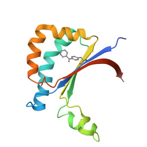
    9IKU, 9ILO, 9IM9, 9IPR

  • PubMed Abstract: 

    The creation of enzymes with abiological abilities offers exciting opportunities to access new-to-nature biocatalysis beyond that found in nature. Here, we repurpose a novel protein scaffold, CTB10, as an artificial photoenzyme through genetic code expansion. It enables catalytic deracemization of cyclopropane, a process that remains inaccessible to traditional biocatalysis due to its thermodynamically unfavorable nature. Following structural optimization through directed evolution, a broad substrate scope with high enantioselectivities is achieved. Furthermore, the crystal structure of the CTB10-based photoenzyme-substrate complex well demonstrates how the catalytic chiral cavity is sculpted to promote efficient and selective light-enabled deracemization. Therefore, this study unlocks the potential for achieving challenging deracemization through biocatalysis.


  • Organizational Affiliation
    • Key Laboratory of Carbohydrate Chemistry and Biotechnology, Ministry of Education, School of Biotechnology, Jiangnan University, Wuxi 214122, P. R. China.

Macromolecules
Find similar proteins by:  (by identity cutoff)  |  3D Structure
Entity ID: 1
MoleculeChains Sequence LengthOrganismDetailsImage
CTB10
A, B, C, D, E
A, B, C, D, E, F, G, H
141Cercospora sp. JNU001Mutation(s): 1 
UniProt
Find proteins for A0A977K7H6 (Cercospora sp. JNU001)
Explore A0A977K7H6 
Go to UniProtKB:  A0A977K7H6
Entity Groups  
Sequence Clusters30% Identity50% Identity70% Identity90% Identity95% Identity100% Identity
UniProt GroupA0A977K7H6
Sequence Annotations
Expand
  • Reference Sequence
Small Molecules
Modified Residues  1 Unique
IDChains TypeFormula2D DiagramParent
PBF
Query on PBF
A, B, C, D, E
A, B, C, D, E, F, G, H
L-PEPTIDE LINKINGC16 H15 N O3PHE
Experimental Data & Validation

Experimental Data

  • Method: X-RAY DIFFRACTION
  • Resolution: 1.94 Å
  • R-Value Free:  0.226 (Depositor), 0.227 (DCC) 
  • R-Value Work:  0.191 (Depositor), 0.194 (DCC) 
  • R-Value Observed: 0.193 (Depositor) 
Space Group: P 1 21 1
Unit Cell:
Length ( Å )Angle ( ˚ )
a = 83.08α = 90
b = 88.114β = 116.66
c = 87.597γ = 90
Software Package:
Software NamePurpose
PHENIXrefinement
xia2data reduction
Aimlessdata scaling
MOLREPphasing

Structure Validation

View Full Validation Report


Entry History & Funding Information

Deposition Data


Funding OrganizationLocationGrant Number
Other privateChina2021YFC2102700
Other privateChinaBK20202002
Other privateChinaKYCX20_1812
Other privateChinaKYCX20_1816

Revision History  (Full details and data files)

  • Version 1.0: 2025-05-14
    Type: Initial release