9J1O | pdb_00009j1o

Mouse Spi-B Ets domain in complex with DNA containing GGAA sequence


Experimental Data Snapshot

  • Method: X-RAY DIFFRACTION
  • Resolution: 2.23 Å
  • R-Value Free: 
    0.275 (Depositor), 0.275 (DCC) 
  • R-Value Work: 
    0.228 (Depositor), 0.227 (DCC) 
  • R-Value Observed: 
    0.230 (Depositor) 

Starting Model: experimental
View more details

wwPDB Validation   3D Report Full Report


Ligand Structure Quality Assessment 


This is version 1.0 of the entry. See complete history


Literature

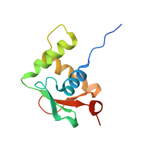
Structural analysis of Spi-B DNA-binding Ets domain recognizing 5'-AGAA-3' and 5'-GGAA-3' sequences.

Nonaka, Y.Hoshino, K.Nakamura, T.Kamitori, S.

(2025) Biochem Biophys Res Commun 749: 151354-151354

  • DOI: https://doi.org/10.1016/j.bbrc.2025.151354
  • Primary Citation of Related Structures:  
    9J1N, 9J1O

  • PubMed Abstract: 

    Plasmacytoid dendritic cells produce large amounts of type-I interferon (IFN-I) upon sensing nucleic acid components of pathogens by Toll-like receptors (TLR7 and TLR9). The transcription factor Spi-B has the DNA-binding Ets domain, and transactivates the Ifna4 promoter co-operatively with IFN regulatory factor-7 (IRF-7) for TLR7/TLR9-induced IFN-I production. Spi-B associates with IRF-7, and activates transcription by binding to the 5'-AGAA-3' sequence, being different from 5'-GGAA-3', known as the Ets domain recognition sequence. To understand the molecular mechanism for the co-operative transactivation of the Ifna4 promoter by Spi-B and IRF-7, we performed X-ray structural determination of the Spi-B Ets domain in complex with target DNAs, including 5'-AGAA-3' and 5'-GGAA-3' sequences. Furthermore, we conducted a modeling study of the complex of the Spi-B and IRF-7 with Ifna4 promoter DNA. X-ray structures showed that the binding of the Spi-B Ets domain induces a kink in DNA at the recognition sequence, and a more kinked DNA structure was observed in 5'-AGAA-3' than 5'-GGAA-3'. A modeling study showed that the Spi-B-induced kinked DNA structure in 5'-AGAA-3' is favorable for Spi-B and IRF-7 to approach each other for association on DNA.


  • Organizational Affiliation

    International Institute of Rare Sugar Research and Education, Kagawa University, Takamatsu, Kagawa, 760-8521, Japan; Department of Pharmacology, Faculty of Medicine, Kagawa University, 1750-1 Ikenobe, Miki-cho, Kita-gun, Kagawa, 761-0793, Japan.


Macromolecules

Find similar proteins by:  (by identity cutoff)  |  3D Structure
Entity ID: 3
MoleculeChains Sequence LengthOrganismDetailsImage
Transcription factor Spi-B106Mus musculusMutation(s): 0 
Gene Names: Spib
UniProt
Find proteins for O35906 (Mus musculus)
Explore O35906 
Go to UniProtKB:  O35906
Entity Groups  
Sequence Clusters30% Identity50% Identity70% Identity90% Identity95% Identity100% Identity
UniProt GroupO35906
Sequence Annotations
Expand
  • Reference Sequence

Find similar nucleic acids by:  Sequence   |   3D Structure  

Entity ID: 1
MoleculeChains LengthOrganismImage
DNA (5'-D(*(3D1)P*TP*GP*AP*AP*AP*AP*GP*GP*GP*AP*AP*TP*TP*GP*G)-3')15Mus musculus
Sequence Annotations
Expand
  • Reference Sequence

Find similar nucleic acids by:  Sequence   |   3D Structure  

Entity ID: 2
MoleculeChains LengthOrganismImage
DNA (5'-D(*(THM)P*CP*CP*AP*AP*TP*TP*CP*CP*CP*TP*TP*TP*TP*CP*A)-3')15Mus musculus
Sequence Annotations
Expand
  • Reference Sequence
Experimental Data & Validation

Experimental Data

  • Method: X-RAY DIFFRACTION
  • Resolution: 2.23 Å
  • R-Value Free:  0.275 (Depositor), 0.275 (DCC) 
  • R-Value Work:  0.228 (Depositor), 0.227 (DCC) 
  • R-Value Observed: 0.230 (Depositor) 
Space Group: P 21 21 21
Unit Cell:
Length ( Å )Angle ( ˚ )
a = 33.2α = 90
b = 54.87β = 90
c = 100.79γ = 90
Software Package:
Software NamePurpose
PHENIXrefinement
XDSdata reduction
XSCALEdata scaling
MOLREPphasing

Structure Validation

View Full Validation Report


Ligand Structure Quality Assessment 


Entry History & Funding Information

Deposition Data


Funding OrganizationLocationGrant Number
Not funded--

Revision History  (Full details and data files)

  • Version 1.0: 2025-06-18
    Type: Initial release