9JAP | pdb_00009jap

Helical structure of EfAvs5(SIR2-STAND)


Experimental Data Snapshot

  • Method: ELECTRON MICROSCOPY
  • Resolution: 3.40 Å
  • Aggregation State: PARTICLE 
  • Reconstruction Method: SINGLE PARTICLE 

wwPDB Validation   3D Report Full Report


This is version 1.1 of the entry. See complete history


Literature

Filamentation activates bacterial Avs5 antiviral protein.

Wang, Y.Tian, Y.Yang, X.Yu, F.Zheng, J.

(2025) Nat Commun 16: 2408-2408

  • DOI: https://doi.org/10.1038/s41467-025-57732-7
  • Primary Citation of Related Structures:  
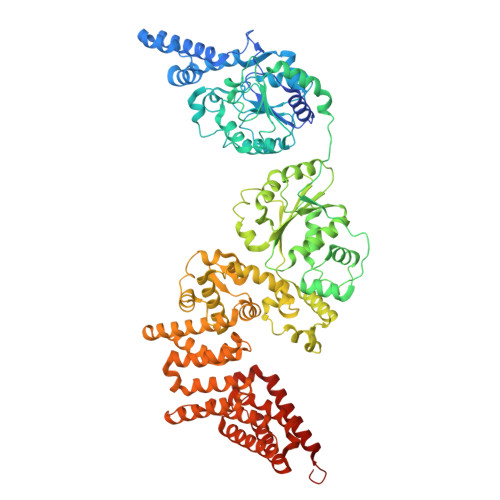
    9JAP

  • PubMed Abstract: 

    Bacterial antiviral STANDs (Avs) are evolutionarily related to the nucleotide-binding oligomerization domain (NOD)-like receptors widely distributed in immune systems across animals and plants. EfAvs5, a type 5 Avs from Escherichia fergusonii, contains an N-terminal SIR2 effector domain, a NOD, and a C-terminal sensor domain, conferring protection against diverse phage invasions. Despite the established roles of SIR2 and STAND in prokaryotic and eukaryotic immunity, the mechanism underlying their collaboration remains unclear. Here we present cryo-EM structures of EfAvs5 filaments, elucidating the mechanisms of dimerization, filamentation, filament bundling, ATP binding, and NAD + hydrolysis, all of which are crucial for anti-phage defense. The SIR2 and NOD domains engage in intra- and inter-dimer interaction to form an individual filament, while the outward C-terminal sensor domains contribute to bundle formation. Filamentation potentially stabilizes the dimeric SIR2 configuration, thereby activating the NADase activity of EfAvs5. Furthermore, we identify the nucleotide kinase gp1.7 of phage T7 as an activator of EfAvs5, demonstrating its ability to induce filamentation and NADase activity. Together, we uncover the filament assembly of Avs5 as a unique mechanism to switch enzyme activities and perform anti-phage defenses.


  • Organizational Affiliation
    • State Key Laboratory of Microbial Metabolism, School of Life Sciences and Biotechnology, Shanghai Jiao Tong University, Shanghai, China.

Macromolecules
Find similar proteins by:  (by identity cutoff)  |  3D Structure
Entity ID: 1
MoleculeChains Sequence LengthOrganismDetailsImage
SIR2-like domain-containing protein
A, B, C, D, E
A, B, C, D, E, F, G, H
869Escherichia fergusoniiMutation(s): 0 
UniProt
Find proteins for A0AA94GZI5 (Kosakonia oryzae)
Explore A0AA94GZI5 
Go to UniProtKB:  A0AA94GZI5
Entity Groups  
Sequence Clusters30% Identity50% Identity70% Identity90% Identity95% Identity100% Identity
UniProt GroupA0AA94GZI5
Sequence Annotations
Expand
  • Reference Sequence
Small Molecules
Ligands 2 Unique
IDChains Name / Formula / InChI Key2D Diagram3D Interactions
ATP (Subject of Investigation/LOI)
Query on ATP

Download Ideal Coordinates CCD File 
I [auth A]
K [auth B]
M [auth C]
O [auth D]
Q [auth E]
I [auth A],
K [auth B],
M [auth C],
O [auth D],
Q [auth E],
S [auth F],
U [auth G],
W [auth H]
ADENOSINE-5'-TRIPHOSPHATE
C10 H16 N5 O13 P3
ZKHQWZAMYRWXGA-KQYNXXCUSA-N
MG (Subject of Investigation/LOI)
Query on MG

Download Ideal Coordinates CCD File 
J [auth A]
L [auth B]
N [auth C]
P [auth D]
R [auth E]
J [auth A],
L [auth B],
N [auth C],
P [auth D],
R [auth E],
T [auth F],
V [auth G],
X [auth H]
MAGNESIUM ION
Mg
JLVVSXFLKOJNIY-UHFFFAOYSA-N
Experimental Data & Validation

Experimental Data

  • Method: ELECTRON MICROSCOPY
  • Resolution: 3.40 Å
  • Aggregation State: PARTICLE 
  • Reconstruction Method: SINGLE PARTICLE 
EM Software:
TaskSoftware PackageVersion
MODEL REFINEMENTPHENIX1.20.1-4487

Structure Validation

View Full Validation Report


Entry History & Funding Information

Deposition Data


Funding OrganizationLocationGrant Number
National Natural Science Foundation of China (NSFC)China31770068, 32070040

Revision History  (Full details and data files)

  • Version 1.0: 2025-03-19
    Type: Initial release
  • Version 1.1: 2025-03-26
    Changes: Data collection, Database references