9OA2 | pdb_00009oa2

Ecoli DnaB helicase and Phage Lambda loader P with ADP-Mg in a 6:6 stoichiometry ratio.


Experimental Data Snapshot

  • Method: ELECTRON MICROSCOPY
  • Resolution: 3.85 Å
  • Aggregation State: PARTICLE 
  • Reconstruction Method: SINGLE PARTICLE 

Starting Models: experimental, in silico
View more details

wwPDB Validation   3D Report Full Report


This is version 1.0 of the entry. See complete history


Literature

Distinct Quaternary States, Intermediates, and Autoinhibition During Loading of the DnaB-Replicative Helicase by the Phage lambda P Helicase Loader.

Shatarupa, A.Brown, D.Olinares, P.D.B.Chase, J.Isiorho, E.Chait, B.T.Jeruzalmi, D.

(2025) bioRxiv 

  • DOI: https://doi.org/10.1101/2022.12.30.522210
  • Primary Citation of Related Structures:  
    8V9S, 9OA1, 9OA2

  • PubMed Abstract: 

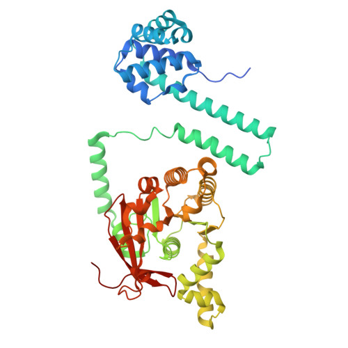
    Replicative helicases require loader proteins for assembly at the origins of DNA replication. Multiple copies of the bacteriophage λP (P) loader bind to and load the E. coli DnaB (B) replicative helicase on replication-origin-derived single-stranded DNA. We find that the E. coli DnaB•λP complex exists in two forms: B 6 P 5 and B 6 P 6 . In the 2.66 Å cryo-EM model of B 6 P 5 , five copies of the λP loader assemble into a crown-like shape that tightly grips DnaB. In this complex, closed planar DnaB is reconfigured into an open spiral with a sufficiently sized breach to permit ssDNA to enter an internal chamber. The transition to the open spiral involves λP-mediated changes to the Docking Helix (DH)-Linker Helix (LH) interface. The loader directly stabilizes the open spiral. Unexpectedly, one λP chain in B 6 P 5 is bound across the breach, precluding entry of replication-origin-derived ssDNA into DnaB's central chamber. We suggest that the B 6 P 6 complex is an early intermediate in the helicase activation pathway wherein neither the DnaB helicase nor the λP loader has attained its final form. DnaB in this complex adopts a partially open planar configuration, termed ajar planar. The partially ordered λP loader assembly features a much looser interaction with DnaB. The ssDNA and ATP sites in both complexes are in a configuration ill-suited for binding or hydrolysis. Our work specifies the conformational changes required for the intermediate B 6 P 6 to transition to B 6 P 5 on the pathway to recruitment by the initiator protein complex to the replication origin.


Macromolecules
Find similar proteins by:  (by identity cutoff)  |  3D Structure
Entity ID: 1
MoleculeChains Sequence LengthOrganismDetailsImage
Replicative DNA helicase
A, B, C, D, E
A, B, C, D, E, F
471Escherichia coliMutation(s): 0 
Gene Names: dnaBgroPgrpAb4052JW4012
EC: 3.6.4.12 (PDB Primary Data), 5.6.2.3 (UniProt)
UniProt
Find proteins for P0ACB0 (Escherichia coli (strain K12))
Explore P0ACB0 
Go to UniProtKB:  P0ACB0
Entity Groups  
Sequence Clusters30% Identity50% Identity70% Identity90% Identity95% Identity100% Identity
UniProt GroupP0ACB0
Sequence Annotations
Expand
  • Reference Sequence
Find similar proteins by:  (by identity cutoff)  |  3D Structure
Entity ID: 2
MoleculeChains Sequence LengthOrganismDetailsImage
Helicase loader233Escherichia phage LambdaMutation(s): 1 
UniProt
Find proteins for P03689 (Escherichia phage lambda)
Explore P03689 
Go to UniProtKB:  P03689
Entity Groups  
Sequence Clusters30% Identity50% Identity70% Identity90% Identity95% Identity100% Identity
UniProt GroupP03689
Sequence Annotations
Expand
  • Reference Sequence
Small Molecules
Ligands 2 Unique
IDChains Name / Formula / InChI Key2D Diagram3D Interactions
ADP (Subject of Investigation/LOI)
Query on ADP

Download Ideal Coordinates CCD File 
M [auth A]
O [auth B]
Q [auth C]
S [auth D]
U [auth E]
M [auth A],
O [auth B],
Q [auth C],
S [auth D],
U [auth E],
W [auth F]
ADENOSINE-5'-DIPHOSPHATE
C10 H15 N5 O10 P2
XTWYTFMLZFPYCI-KQYNXXCUSA-N
MG (Subject of Investigation/LOI)
Query on MG

Download Ideal Coordinates CCD File 
N [auth A]
P [auth B]
R [auth C]
T [auth D]
V [auth E]
N [auth A],
P [auth B],
R [auth C],
T [auth D],
V [auth E],
X [auth F]
MAGNESIUM ION
Mg
JLVVSXFLKOJNIY-UHFFFAOYSA-N
Experimental Data & Validation

Experimental Data

  • Method: ELECTRON MICROSCOPY
  • Resolution: 3.85 Å
  • Aggregation State: PARTICLE 
  • Reconstruction Method: SINGLE PARTICLE 
EM Software:
TaskSoftware PackageVersion
RECONSTRUCTIONcryoSPARC4
MODEL REFINEMENTPHENIX1.21.2_5419

Structure Validation

View Full Validation Report


Entry History & Funding Information

Deposition Data


Funding OrganizationLocationGrant Number
National Science Foundation (NSF, United States)United States1818255

Revision History  (Full details and data files)

  • Version 1.0: 2025-07-16
    Type: Initial release