9OH6 | pdb_00009oh6

H117A/M121H Azurin with Cu(II), pH 7.7


Experimental Data Snapshot

  • Method: X-RAY DIFFRACTION
  • Resolution: 2.04 Å
  • R-Value Free: 
    0.233 (Depositor), 0.233 (DCC) 
  • R-Value Work: 
    0.176 (Depositor), 0.177 (DCC) 
  • R-Value Observed: 
    0.179 (Depositor) 

Starting Model: experimental
View more details

wwPDB Validation   3D Report Full Report


This is version 1.0 of the entry. See complete history


Literature

Beyond Blue: Systematic Modulation of Electronic Structure and Redox Properties of Type 1 Copper in Azurin.

Van Stappen, C.Xu, J.Liu, Y.Van Stappen, J.Kim, W.Zhang, Y.J.Lu, Y.

(2025) J Am Chem Soc 147: 24825-24837

  • DOI: https://doi.org/10.1021/jacs.5c07009
  • Primary Citation of Related Structures:  
    9OH6, 9OH7

  • PubMed Abstract: 

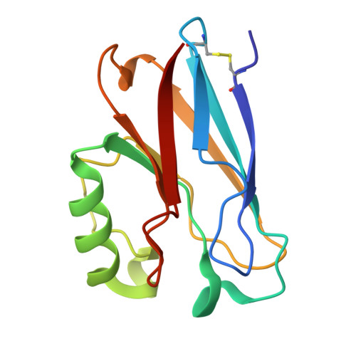
    The reduction potentials of metal ions ( E °'), crucial for optimizing biological processes like electron transfer and catalysis, are finely tuned by interactions between the primary and secondary coordination spheres (PCS, SCS). While previous successes in tuning E °' in azurin have provided deeper insights into how the SCS influences electronic structure and associated redox properties of "classic" blue copper proteins, our understanding of E °' tuning in other subclasses of type 1 Cu (T1Cu) proteins, such as green and red copper proteins, remains rudimentary. To address this issue, we report the design of a green copper center in azurin where an equatorial-to-axial shift in a histidine binding interaction leads to reorientation of the Cu-centered redox active molecular orbital and a +100 mV shift in E °'. In contrast to a 22 mV decrease in E °' when a hydrophobic interaction is introduced in wild-type azurin through the Met13Phe mutation, this same mutation leads to a 65 mV increase in our designed green Cu azurin. More importantly, using a combination of EPR spectroscopy, protein crystallography, and quantum mechanical calculations, we uncover correlations between E °', d-s orbital mixing, and the angle between S Cys -Cu and N δH46 -Cu bonds, ∠(S Cys -Cu-N δH46 ), allowing rationalization of increases in E °' of green Cu proteins through an entropically driven T-shape distortion. By providing direct connections between geometry, electronic structure, and functional properties such as E °', this work opens previously unexplored routes to systematically modulating E °' through the combination of spatial reorientation of the redox active molecular orbital and varying geometric distortion in the primary coordination sphere.


  • Organizational Affiliation

    Department of Chemistry, University of Texas at Austin, 105 E. 24th Street, Austin, Texas 78712, United States.


Macromolecules
Find similar proteins by:  (by identity cutoff)  |  3D Structure
Entity ID: 1
MoleculeChains Sequence LengthOrganismDetailsImage
AzurinA [auth B],
B [auth E],
C [auth H],
D [auth K]
128Pseudomonas aeruginosa PAO1Mutation(s): 2 
Gene Names: azuPA4922
UniProt
Find proteins for P00282 (Pseudomonas aeruginosa (strain ATCC 15692 / DSM 22644 / CIP 104116 / JCM 14847 / LMG 12228 / 1C / PRS 101 / PAO1))
Explore P00282 
Go to UniProtKB:  P00282
Entity Groups  
Sequence Clusters30% Identity50% Identity70% Identity90% Identity95% Identity100% Identity
UniProt GroupP00282
Sequence Annotations
Expand
  • Reference Sequence
Small Molecules
Ligands 2 Unique
IDChains Name / Formula / InChI Key2D Diagram3D Interactions
TRS
Query on TRS

Download Ideal Coordinates CCD File 
E [auth B],
H [auth E],
K [auth H],
N [auth K]
2-AMINO-2-HYDROXYMETHYL-PROPANE-1,3-DIOL
C4 H12 N O3
LENZDBCJOHFCAS-UHFFFAOYSA-O
CU (Subject of Investigation/LOI)
Query on CU

Download Ideal Coordinates CCD File 
F [auth B]
G [auth B]
I [auth E]
J [auth E]
L [auth H]
F [auth B],
G [auth B],
I [auth E],
J [auth E],
L [auth H],
M [auth H],
O [auth K],
P [auth K]
COPPER (II) ION
Cu
JPVYNHNXODAKFH-UHFFFAOYSA-N
Experimental Data & Validation

Experimental Data

  • Method: X-RAY DIFFRACTION
  • Resolution: 2.04 Å
  • R-Value Free:  0.233 (Depositor), 0.233 (DCC) 
  • R-Value Work:  0.176 (Depositor), 0.177 (DCC) 
  • R-Value Observed: 0.179 (Depositor) 
Space Group: P 1 21 1
Unit Cell:
Length ( Å )Angle ( ˚ )
a = 58.905α = 90
b = 48.836β = 90
c = 81.31γ = 90
Software Package:
Software NamePurpose
PHENIXrefinement
DIALSdata reduction
DIALSdata scaling
PHENIXphasing

Structure Validation

View Full Validation Report


Entry History & Funding Information

Deposition Data


Funding OrganizationLocationGrant Number
National Science Foundation (NSF, United States)United StatesCHE-2420683
National Institutes of Health/National Institute of General Medical Sciences (NIH/NIGMS)United StatesF32GM145134
Robert A. Welch FoundationUnited StatesF-0020

Revision History  (Full details and data files)

  • Version 1.0: 2025-07-23
    Type: Initial release