2YZC | pdb_00002yzc

Crystal structure of uricase from Arthrobacter globiformis in complex with allantoate


Experimental Data Snapshot

  • Method: X-RAY DIFFRACTION
  • Resolution: 1.88 Å
  • R-Value Free: 
    0.195 (Depositor), 0.200 (DCC) 
  • R-Value Work: 
    0.159 (Depositor), 0.180 (DCC) 
  • R-Value Observed: 
    0.161 (Depositor) 

Starting Model: experimental
View more details

wwPDB Validation   3D Report Full Report


Ligand Structure Quality Assessment 


This is version 1.3 of the entry. See complete history


Literature

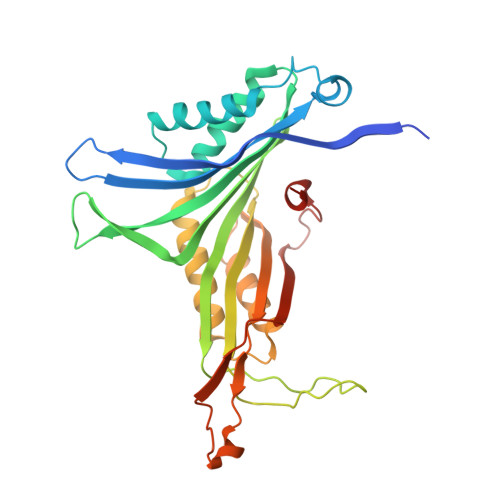
Structures of Arthrobacter globiformis urate oxidase-ligand complexes.

Juan, E.C.Hoque, M.M.Shimizu, S.Hossain, M.T.Yamamoto, T.Imamura, S.Suzuki, K.Tsunoda, M.Amano, H.Sekiguchi, T.Takenaka, A.

(2008) Acta Crystallogr D Biol Crystallogr D64: 815-822

  • DOI: https://doi.org/10.1107/S0907444908013590
  • Primary Citation of Related Structures:  
    2YZB, 2YZC, 2YZD, 2YZE

  • PubMed Abstract: 

    The enzyme urate oxidase catalyzes the conversion of uric acid to 5-hydroxyisourate, one of the steps in the ureide pathway. Arthrobacter globiformis urate oxidase (AgUOX) was crystallized and structures of crystals soaked in the substrate uric acid, the inhibitor 8-azaxanthin and allantoin have been determined at 1.9-2.2 A resolution. The biological unit is a homotetramer and two homotetramers comprise the asymmetric crystallographic unit. Each subunit contains two T-fold domains of betabetaalphaalphabetabeta topology, which are usually found in purine- and pterin-binding enzymes. The uric acid substrate is bound tightly to the enzyme by interactions with Arg180, Leu222 and Gln223 from one subunit and with Thr67 and Asp68 of the neighbouring subunit in the tetramer. In the other crystal structures, lithium borate, 8-azaxanthin and allantoate are bound to the enzyme in a similar manner as uric acid. Based on these AgUOX structures, the enzymatic reaction mechanism of UOX has been proposed.


  • Organizational Affiliation
    • Graduate School of Bioscience and Biotechnology, Tokyo Institute of Technology, 4259 Nagatsuta, Midori-ku, Yokohama, Kanagawa 226-8501, Japan.

Macromolecules
Find similar proteins by:  (by identity cutoff)  |  3D Structure
Entity ID: 1
MoleculeChains Sequence LengthOrganismDetailsImage
Uricase
A, B, C, D, E
A, B, C, D, E, F, G, H
302Arthrobacter globiformisMutation(s): 0 
EC: 1.7.3.3
UniProt
Find proteins for D0VWQ1 (Arthrobacter globiformis)
Explore D0VWQ1 
Go to UniProtKB:  D0VWQ1
Entity Groups  
Sequence Clusters30% Identity50% Identity70% Identity90% Identity95% Identity100% Identity
UniProt GroupD0VWQ1
Sequence Annotations
Expand
  • Reference Sequence
Experimental Data & Validation

Experimental Data

  • Method: X-RAY DIFFRACTION
  • Resolution: 1.88 Å
  • R-Value Free:  0.195 (Depositor), 0.200 (DCC) 
  • R-Value Work:  0.159 (Depositor), 0.180 (DCC) 
  • R-Value Observed: 0.161 (Depositor) 
Space Group: P 21 21 21
Unit Cell:
Length ( Å )Angle ( ˚ )
a = 85.498α = 90
b = 122.942β = 90
c = 284.784γ = 90
Software Package:
Software NamePurpose
REFMACrefinement
HKL-2000data collection
HKL-2000data reduction
HKL-2000data scaling
AMoREphasing

Structure Validation

View Full Validation Report


Ligand Structure Quality Assessment 


Entry History 

Deposition Data

Revision History  (Full details and data files)

  • Version 1.0: 2008-05-06
    Type: Initial release
  • Version 1.1: 2011-07-13
    Changes: Version format compliance
  • Version 1.2: 2023-10-25
    Changes: Data collection, Database references, Derived calculations, Refinement description
  • Version 1.3: 2025-08-06
    Changes: Database references, Structure summary