8XR5 | pdb_00008xr5

Crystal structure of PD-L1 complexed with small molecule inhibitor X18


Experimental Data Snapshot

  • Method: X-RAY DIFFRACTION
  • Resolution: 1.95 Å
  • R-Value Free: 
    0.257 (Depositor), 0.233 (DCC) 
  • R-Value Work: 
    0.227 (Depositor), 0.230 (DCC) 
  • R-Value Observed: 
    0.230 (Depositor) 

Starting Model: experimental
View more details

wwPDB Validation   3D Report Full Report


Ligand Structure Quality Assessment 


This is version 1.0 of the entry. See complete history


Literature

Discovery of Novel PD-L1 Small-Molecular Inhibitors with Potent In Vivo Anti-tumor Immune Activity.

Liu, L.Zhang, H.Hou, J.Zhang, Y.Wang, L.Wang, S.Yao, Z.Xie, T.Wen, X.Xu, Q.Dai, L.Feng, Z.Zhang, P.Wu, Y.Sun, H.Liu, J.Yuan, H.

(2024) J Med Chem 67: 4977-4997

  • DOI: https://doi.org/10.1021/acs.jmedchem.4c00102
  • Primary Citation of Related Structures:  
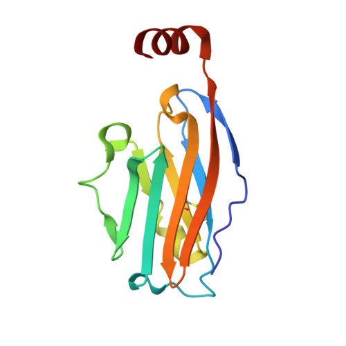
    8XR5

  • PubMed Abstract: 

    Programmed death-ligand 1 (PD-L1) has surfaced as a promising therapeutic target for various cancers due to its pivotal role in facilitating tumor immune evasion. Herein, we report a series of novel small-molecule PD-L1 inhibitors exhibiting remarkable inhibitory activity against the PD-1/PD-L1 interaction ( X18 : IC 50 = 1.3 nM) and reinstating the suppressive effect of PD-L1 on T cells ( X18 : EC 50 = 152.8 nM). Crystallographic studies revealed the binding mode of X18 and PD-L1. Through a rational prodrug design approach, we have successfully optimized the oral pharmacokinetic properties of X22 , effectively addressing the poor oral pharmacokinetic profile of PD-L1 small-molecule inhibitors. Notably, X22 demonstrated significant antitumor efficacy in murine models of MC38 and CT26 colon cancer through the upregulation of tumor infiltration and cytotoxicity of CD8 + T cells partially. These findings offer promising prospects for the advancement of PD-L1 inhibitors as innovative agents in cancer immunotherapy.


  • Organizational Affiliation

    Jiangsu Key Laboratory of Drug Discovery for Metabolic Disease, State Key Laboratory of Natural Medicines, China Pharmaceutical University, Nanjing 210009, China.


Macromolecules
Find similar proteins by:  (by identity cutoff)  |  3D Structure
Entity ID: 1
MoleculeChains Sequence LengthOrganismDetailsImage
Programmed cell death 1 ligand 1
A, B
126Homo sapiensMutation(s): 0 
Gene Names: CD274B7H1PDCD1L1PDCD1LG1PDL1
UniProt & NIH Common Fund Data Resources
Find proteins for Q9NZQ7 (Homo sapiens)
Explore Q9NZQ7 
Go to UniProtKB:  Q9NZQ7
PHAROS:  Q9NZQ7
GTEx:  ENSG00000120217 
Entity Groups  
Sequence Clusters30% Identity50% Identity70% Identity90% Identity95% Identity100% Identity
UniProt GroupQ9NZQ7
Sequence Annotations
Expand
  • Reference Sequence
Small Molecules
Ligands 1 Unique
IDChains Name / Formula / InChI Key2D Diagram3D Interactions
A1LV3
Query on A1LV3

Download Ideal Coordinates CCD File 
C [auth B](2~{R})-2-[[2-(2,1,3-benzoxadiazol-5-ylmethoxy)-5-chloranyl-4-[[2-fluoranyl-3-[3-[3-(4-oxidanylpiperidin-1-yl)propoxy]phenyl]phenyl]methoxy]phenyl]methylamino]-3-oxidanyl-propanoic acid
C38 H40 Cl F N4 O8
CVHGLSHXHMFDJX-UUWRZZSWSA-N
Experimental Data & Validation

Experimental Data

  • Method: X-RAY DIFFRACTION
  • Resolution: 1.95 Å
  • R-Value Free:  0.257 (Depositor), 0.233 (DCC) 
  • R-Value Work:  0.227 (Depositor), 0.230 (DCC) 
  • R-Value Observed: 0.230 (Depositor) 
Space Group: P 21 21 2
Unit Cell:
Length ( Å )Angle ( ˚ )
a = 109.171α = 90
b = 49.09β = 90
c = 51.849γ = 90
Software Package:
Software NamePurpose
PHENIXrefinement
HKL-2000data scaling
HKL-2000data reduction
PHENIXphasing

Structure Validation

View Full Validation Report


Ligand Structure Quality Assessment 


Entry History & Funding Information

Deposition Data

  • Released Date: 2025-02-26 
  • Deposition Author(s): Liu, L.

Funding OrganizationLocationGrant Number
National Natural Science Foundation of China (NSFC)China82204216

Revision History  (Full details and data files)

  • Version 1.0: 2025-02-26
    Type: Initial release