9MGB | pdb_00009mgb

scFv antibody CL33 bound to R-phycoerythrin


Experimental Data Snapshot

  • Method: ELECTRON MICROSCOPY
  • Resolution: 2.10 Å
  • Aggregation State: PARTICLE 
  • Reconstruction Method: SINGLE PARTICLE 

wwPDB Validation   3D Report Full Report


This is version 1.1 of the entry. See complete history


Literature

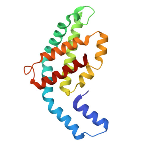
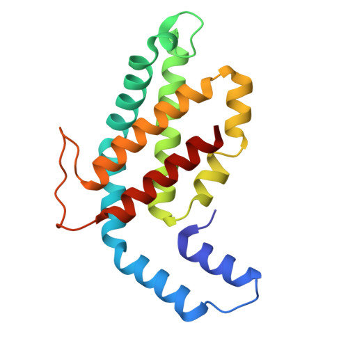
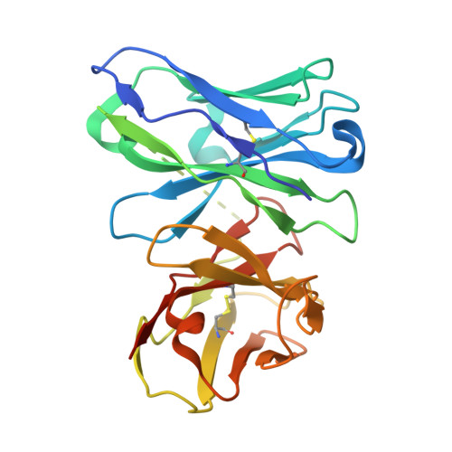
Antibody-like recognition of a gamma delta T cell receptor toward a foreign antigen.

Rashleigh, L.Venugopal, H.Rice, M.T.Gunasinghe, S.D.Sok, C.L.Gherardin, N.A.Almeida, C.F.Van Rhijn, I.Moody, D.B.Godfrey, D.I.Rossjohn, J.Gully, B.S.

(2025) Structure 

  • DOI: https://doi.org/10.1016/j.str.2025.07.006
  • Primary Citation of Related Structures:  
    9MGB, 9MKO, 9O60, 9O61, 9O62

  • PubMed Abstract: 

    The antigen recognition principles of B cells and αβ T cells have been well described compared to those of the γδ T cell. By way of their specificity conferring receptor (γδTCR), γδ T cells can directly bind proteinaceous antigens. A known γδ T cell and B cell model antigen is phycoerythrin (PE), a light harvesting protein from rhodophytes and cyanobacteria. Here we probed human γδTCR reactivity to PE, in which a Vδ1Vγ5 TCR bound directly to induce proximal signaling and cellular activation. We determined the cryoelectron microscopy (cryo-EM) structure of the γδTCR-phycoerythrin immune complex. We then determined the cryo-EM structures of an antibody fragment and an αβTCR bound to PE. This revealed convergent use of apical aromatic residues to mediate contacts with a common PE epitope. Comparative analyses of the γδTCR revealed multiple antibody-like characteristics, including an enrichment of apical aromatic residues. Our findings reveal further distinct facets of antigen recognition by the γδTCR.


  • Organizational Affiliation
    • Infection and Immunity Program & Department of Biochemistry and Molecular Biology, Biomedicine Discovery Institute, Monash University, Clayton, VIC, Australia.

Macromolecules
Find similar proteins by:  (by identity cutoff)  |  3D Structure
Entity ID: 1
MoleculeChains Sequence LengthOrganismDetailsImage
R-phycoerythrin alpha chain164Pyropia teneraMutation(s): 0 
UniProt
Find proteins for O36005 (Griffithsia monilis)
Explore O36005 
Go to UniProtKB:  O36005
Entity Groups  
Sequence Clusters30% Identity50% Identity70% Identity90% Identity95% Identity100% Identity
UniProt GroupO36005
Sequence Annotations
Expand
  • Reference Sequence
Find similar proteins by:  (by identity cutoff)  |  3D Structure
Entity ID: 2
MoleculeChains Sequence LengthOrganismDetailsImage
R-phycoerythrin beta chain176Pyropia teneraMutation(s): 0 
UniProt
Find proteins for P68939 (Pyropia tenera)
Explore P68939 
Go to UniProtKB:  P68939
Entity Groups  
Sequence Clusters30% Identity50% Identity70% Identity90% Identity95% Identity100% Identity
UniProt GroupP68939
Sequence Annotations
Expand
  • Reference Sequence
Find similar proteins by:  (by identity cutoff)  |  3D Structure
Entity ID: 3
MoleculeChains Sequence LengthOrganismDetailsImage
CL33 scFv257Mus musculusMutation(s): 0 
Entity Groups  
Sequence Clusters30% Identity50% Identity70% Identity90% Identity95% Identity100% Identity
Sequence Annotations
Expand
  • Reference Sequence
Small Molecules
Ligands 2 Unique
IDChains Name / Formula / InChI Key2D Diagram3D Interactions
PUB (Subject of Investigation/LOI)
Query on PUB

Download Ideal Coordinates CCD File 
EA [auth G]
JA [auth J]
OA [auth M]
TA [auth Q]
U [auth B]
EA [auth G],
JA [auth J],
OA [auth M],
TA [auth Q],
U [auth B],
Z [auth D]
PHYCOUROBILIN
C33 H42 N4 O6
KDCCOOGTVSRCHX-YYVBKQGDSA-N
PEB (Subject of Investigation/LOI)
Query on PEB

Download Ideal Coordinates CCD File 
AA [auth D]
BA [auth D]
CA [auth F]
DA [auth F]
FA [auth G]
AA [auth D],
BA [auth D],
CA [auth F],
DA [auth F],
FA [auth G],
GA [auth G],
HA [auth I],
IA [auth I],
KA [auth J],
LA [auth J],
MA [auth L],
NA [auth L],
PA [auth M],
QA [auth M],
RA [auth O],
S [auth A],
SA [auth O],
T [auth A],
UA [auth Q],
V [auth B],
VA [auth Q],
W [auth B],
X [auth C],
Y [auth C]
PHYCOERYTHROBILIN
C33 H40 N4 O6
NKCBCVIFPXGHAV-WAVSMFBNSA-N
Experimental Data & Validation

Experimental Data

  • Method: ELECTRON MICROSCOPY
  • Resolution: 2.10 Å
  • Aggregation State: PARTICLE 
  • Reconstruction Method: SINGLE PARTICLE 
EM Software:
TaskSoftware PackageVersion
MODEL REFINEMENTPHENIX1.21.1_5286:

Structure Validation

View Full Validation Report


Entry History & Funding Information

Deposition Data


Funding OrganizationLocationGrant Number
Australian Research Council (ARC)AustraliaDP230102073

Revision History  (Full details and data files)

  • Version 1.0: 2025-08-06
    Type: Initial release
  • Version 1.1: 2025-08-13
    Changes: Data collection, Database references