1DMB

REFINED 1.8 ANGSTROMS STRUCTURE REVEALS THE MECHANISM OF BINDING OF A CYCLIC SUGAR, BETA-CYCLODEXTRIN, TO THE MALTODEXTRIN BINDING PROTEIN


Experimental Data Snapshot

  • Method: X-RAY DIFFRACTION
  • Resolution: 1.80 Å
  • R-Value Work: 0.210 
  • R-Value Observed: 0.210 

wwPDB Validation   3D Report Full Report


This is version 2.1 of the entry. See complete history


Literature

Refined 1.8-A structure reveals the mode of binding of beta-cyclodextrin to the maltodextrin binding protein.

Sharff, A.J.Rodseth, L.E.Quiocho, F.A.

(1993) Biochemistry 32: 10553-10559

  • DOI: https://doi.org/10.1021/bi00091a004
  • Primary Citation of Related Structures:  
    1DMB

  • PubMed Abstract: 

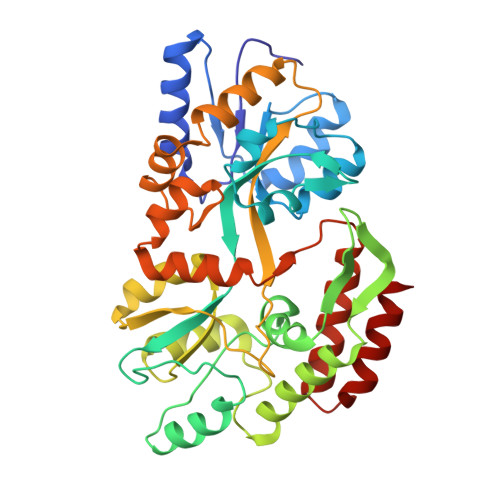
    The maltodextrin binding protein from Escherichia coli serves as the initial receptor for both the active transport of and chemotaxis toward a range of linear maltose sugars. The X-ray structures of both the maltose-bound and sugar-free forms of the protein have been previously described [Spurlino, J. C., Lu, G.-Y., & Quiocho, F. A. (1991) J. Biol. Chem. 266, 5202-5219; Sharff, A. J., Rodseth, L. E., Spurlino, J. C., & Quocho, F. A. (1992) Biochemistry 31, 10657-10663]. The X-ray crystal structure of the maltodextrin binding protein complexed with cyclomaltoheptaose (beta-cyclodextrin) has been determined from a single crystal. The structure has been refined to a final R-value of 21% at 1.8-A resolution. Although not a physiological ligand for the maltodextrin binding protein, beta-cyclodextrin has been shown to bind with a Kd of the same order as those of the linear maltodextrin substrates. The observed structure shows that the complexed protein remains in the fully open conformation and is almost identical to the structure of the unliganded protein. The sugar sits in the open cleft with three glucosyl units bound to the C-domain at the base of the cleft, in a similar position to maltotriose, the most tightly bound ligand. The top of the ring is loosely bound to the upper edge of the cleft on the N-domain. The sugar makes a total of 94 productive interactions (of less than 4.0-A length) with the protein and with bound water molecules.(ABSTRACT TRUNCATED AT 250 WORDS)


  • Organizational Affiliation

    Howard Hughes Medical Institute, Baylor College of Medicine, Houston, Texas 77030.


Macromolecules
Find similar proteins by:  (by identity cutoff)  |  3D Structure
Entity ID: 1
MoleculeChains Sequence LengthOrganismDetailsImage
D-MALTODEXTRIN BINDING PROTEIN370Escherichia coliMutation(s): 0 
UniProt
Find proteins for P0AEX9 (Escherichia coli (strain K12))
Explore P0AEX9 
Go to UniProtKB:  P0AEX9
Entity Groups  
Sequence Clusters30% Identity50% Identity70% Identity90% Identity95% Identity100% Identity
UniProt GroupP0AEX9
Sequence Annotations
Expand
  • Reference Sequence
Oligosaccharides

Help

Entity ID: 2
MoleculeChains Length2D Diagram Glycosylation3D Interactions
Cycloheptakis-(1-4)-(alpha-D-glucopyranose)
B
7N/A
Glycosylation Resources
GlyTouCan:  G01435GL
GlyCosmos:  G01435GL
Biologically Interesting Molecules (External Reference) 1 Unique
Experimental Data & Validation

Experimental Data

  • Method: X-RAY DIFFRACTION
  • Resolution: 1.80 Å
  • R-Value Work: 0.210 
  • R-Value Observed: 0.210 
  • Space Group: P 1
Unit Cell:
Length ( Å )Angle ( ˚ )
a = 38.86α = 101.5
b = 44.32β = 99.3
c = 58.31γ = 102.2
Software Package:
Software NamePurpose
PROLSQrefinement
X-PLORrefinement

Structure Validation

View Full Validation Report


Entry History 

Deposition Data

Revision History  (Full details and data files)

  • Version 1.0: 1993-10-31
    Type: Initial release
  • Version 1.1: 2008-03-24
    Changes: Version format compliance
  • Version 1.2: 2011-07-13
    Changes: Version format compliance
  • Version 1.3: 2019-07-17
    Changes: Data collection, Other, Refinement description
  • Version 1.4: 2019-08-14
    Changes: Data collection, Refinement description
  • Version 2.0: 2020-07-29
    Type: Remediation
    Reason: Carbohydrate remediation
    Changes: Atomic model, Data collection, Derived calculations, Non-polymer description, Structure summary
  • Version 2.1: 2024-02-07
    Changes: Data collection, Database references, Structure summary