Crystal Structure of a Complex Formed between a Snake Venom Phospholipase A2 and a Potent Peptide Inhibitor Phe-Leu-Ser-Tyr-Lys at 1.8 A Resolution
Chandra, V., Jasti, J., Kaur, P., Dey, S., Perbandt, M., Srinivasan, A., Betzel, C., Singh, T.P.(2002) J Biol Chem 277: 41079-41085
- PubMed: 12186870
- DOI: https://doi.org/10.1074/jbc.M206130200
- Primary Citation of Related Structures:
1JQ9 - PubMed Abstract:
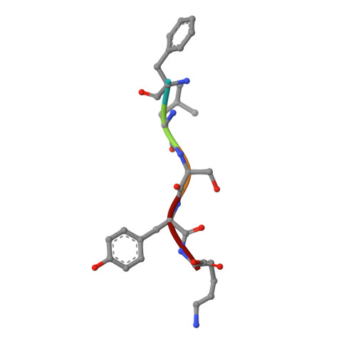
Phospholipase A(2) is an important enzyme involved in the production of prostaglandins and their related compounds causing inflammatory disorders. Among the several peptides tested, the peptide Phe-Leu-Ser-Tyr-Lys (FLSYK) showed the highest inhibition. The dissociation constant (K(d)) for this peptide was calculated to be 3.57 +/- 0.05 x 10(-9) m. In order to further improve the degree of inhibition of phospholipase A(2), a complex between Russells viper snake venom phospholipase A(2) and a peptide inhibitor FLSYK was crystallized, and its structure was determined by crystallographic methods and refined to an R-factor of 0.205 at 1.8 A resolution. The structure contains two crystallographically independent molecules of phospholipase A(2) (molecules A and B) and a peptide molecule specifically bound to molecule A only. The two molecules formed an asymmetric dimer. The dimerization caused a modification in the binding site of molecule A. The overall conformations of molecules A and B were found to be generally similar except three regions i.e. the Trp-31-containing loop (residues 25-34), the beta-wing consisting of two antiparallel beta-strands (residues 74-85) and the C-terminal region (residues 119-133). Out of the above three, the most striking difference pertains to the conformation of Trp-31 in the two molecules. The orientation of Trp-31 in molecule A was suitable for the binding of FLSYK, while it disallowed the binding of peptide to molecule B. The structure of the complex clearly shows that the peptide is so placed in the binding site of molecule A that the side chain of its lysine residue interacted extensively with the enzyme and formed several hydrogen bonds in addition to a strong electrostatic interaction with critical Asp-49. The C-terminal carboxylic group of the peptide interacted with the catalytic residue His-48.
Organizational Affiliation:
Department of Biophysics, All India Institute of Medical Sciences, Ansari Nagar, New Delhi-110029, India.