1Q3U

Crystal structure of a wild-type Cre recombinase-loxP synapse: pre-cleavage complex


Experimental Data Snapshot

  • Method: X-RAY DIFFRACTION
  • Resolution: 2.90 Å
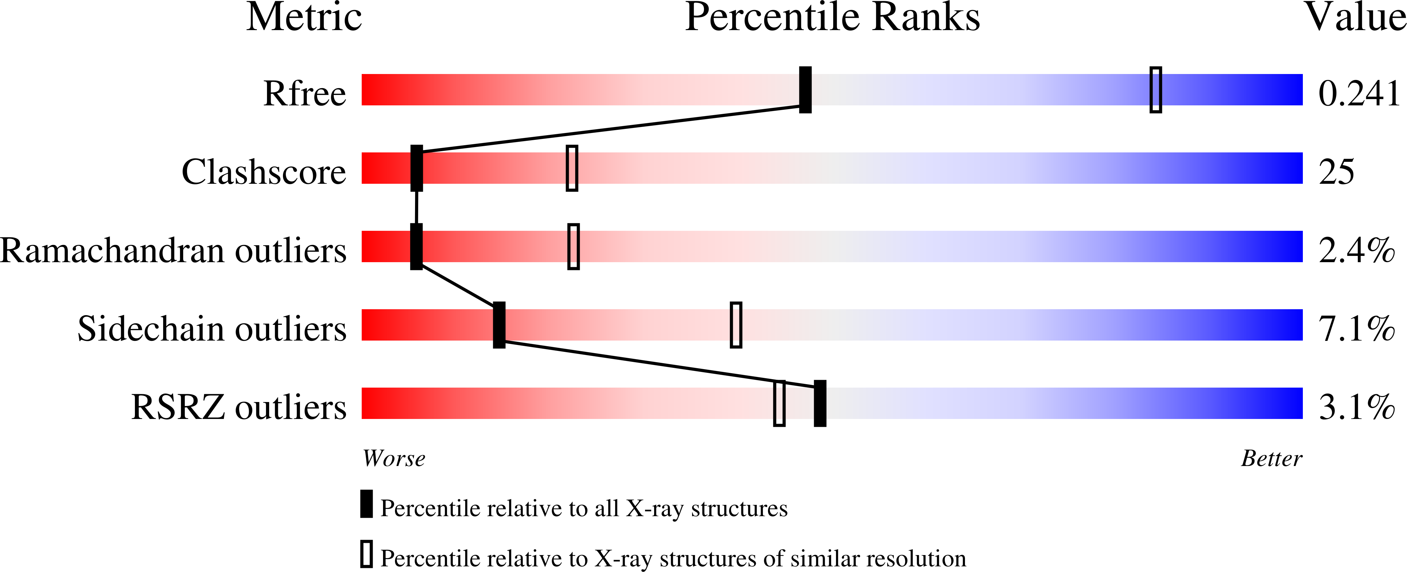
  • R-Value Free: 0.244 
  • R-Value Work: 0.223 
  • R-Value Observed: 0.223 

wwPDB Validation   3D Report Full Report


This is version 1.3 of the entry. See complete history


Literature

Crystal structure of a wild-type Cre recombinase-loxP synapse reveals a novel spacer conformation suggesting an alternative mechanism for DNA cleavage activation

Ennifar, E.Meyer, J.E.W.Buchholz, F.Stewart, A.F.Suck, D.

(2003) Nucleic Acids Res 31: 5449-5460

  • DOI: https://doi.org/10.1093/nar/gkg732
  • Primary Citation of Related Structures:  
    1NZB, 1OUQ, 1Q3U, 1Q3V

  • PubMed Abstract: 

    Escherichia coli phage P1 Cre recombinase catalyzes the site-specific recombination of DNA containing loxP sites. We report here two crystal structures of a wild-type Cre recombinase-loxP synaptic complex corresponding to two distinct reaction states: an initial pre-cleavage complex, trapped using a phosphorothioate modification at the cleavable scissile bond that prevents the recombination reaction, and a 3'-phosphotyrosine protein-DNA intermediate resulting from the first strand cleavage. In contrast to previously determined Cre complexes, both structures contain a full tetrameric complex in the asymmetric unit, unequivocally showing that the anti-parallel arrangement of the loxP sites is an intrinsic property of the Cre-loxP recombination synapse. The conformation of the spacer is different to the one observed for the symmetrized loxS site: a kink next to the scissile phosphate in the top strand of the pre-cleavage complex leads to unstacking of the TpG step and a widening of the minor groove. This side of the spacer is interacting with a 'cleavage-competent' Cre subunit, suggesting that the first cleavage occurs at the ApT step in the top strand. This is further confirmed by the structure of the 3'-phosphotyrosine intermediate, where the DNA is cleaved in the top strands and covalently linked to the 'cleavage-competent' subunits. The cleavage is followed by a movement of the C-terminal part containing the attacking Y324 and the helix N interacting with the 'non-cleaving' subunit. This rearrangement could be responsible for the interconversion of Cre subunits. Our results also suggest that the Cre-induced kink next to the scissile phosphodiester activates the DNA for cleavage at this position and facilitates strand transfer.


  • Organizational Affiliation

    Structural and Computational Biology Programme, EMBL, Meyerhofstrasse 1, D-69117 Heidelberg, Germany.


Macromolecules

Find similar proteins by:  (by identity cutoff)  |  3D Structure
Entity ID: 3
MoleculeChains Sequence LengthOrganismDetailsImage
Cre recombinaseE [auth A],
F [auth B],
G [auth E],
H [auth F]
347Punavirus P1Mutation(s): 0 
UniProt
Find proteins for P06956 (Escherichia phage P1)
Explore P06956 
Go to UniProtKB:  P06956
Entity Groups  
Sequence Clusters30% Identity50% Identity70% Identity90% Identity95% Identity100% Identity
UniProt GroupP06956
Sequence Annotations
Expand
  • Reference Sequence
Find similar nucleic acids by:  (by identity cutoff)  |  3D Structure
Entity ID: 1
MoleculeChains LengthOrganismImage
loxP DNAA [auth C],
C [auth G]
37N/A
Sequence Annotations
Expand
  • Reference Sequence
Find similar nucleic acids by:  (by identity cutoff)  |  3D Structure
Entity ID: 2
MoleculeChains LengthOrganismImage
loxP DNAB [auth D],
D [auth H]
37N/A
Sequence Annotations
Expand
  • Reference Sequence
Small Molecules
Ligands 2 Unique
IDChains Name / Formula / InChI Key2D Diagram3D Interactions
IOD
Query on IOD

Download Ideal Coordinates CCD File 
I [auth C],
S [auth F]
IODIDE ION
I
XMBWDFGMSWQBCA-UHFFFAOYSA-M
MG
Query on MG

Download Ideal Coordinates CCD File 
J [auth C]
K [auth D]
L [auth D]
M [auth G]
N [auth H]
J [auth C],
K [auth D],
L [auth D],
M [auth G],
N [auth H],
O [auth A],
P [auth A],
Q [auth A],
R [auth A],
T [auth F]
MAGNESIUM ION
Mg
JLVVSXFLKOJNIY-UHFFFAOYSA-N
Experimental Data & Validation

Experimental Data

  • Method: X-RAY DIFFRACTION
  • Resolution: 2.90 Å
  • R-Value Free: 0.244 
  • R-Value Work: 0.223 
  • R-Value Observed: 0.223 
  • Space Group: P 21 21 21
Unit Cell:
Length ( Å )Angle ( ˚ )
a = 107.991α = 90
b = 161.064β = 90
c = 196.003γ = 90
Software Package:
Software NamePurpose
DENZOdata reduction
SCALEPACKdata scaling
AMoREphasing
CNSrefinement

Structure Validation

View Full Validation Report


Entry History 

Deposition Data

Revision History  (Full details and data files)

  • Version 1.0: 2003-09-16
    Type: Initial release
  • Version 1.1: 2008-04-29
    Changes: Version format compliance
  • Version 1.2: 2011-07-13
    Changes: Version format compliance
  • Version 1.3: 2023-08-16
    Changes: Data collection, Database references, Derived calculations, Refinement description