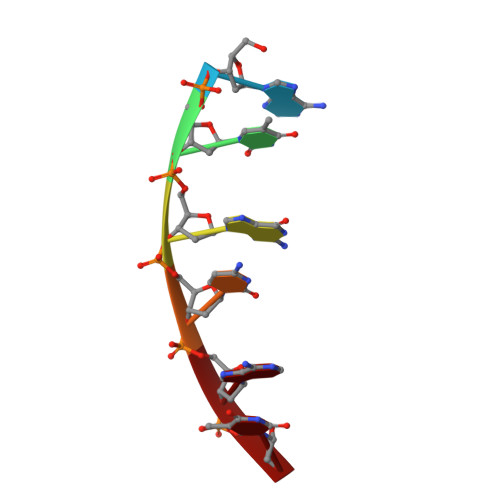
Novel DNA bis-intercalation by MLN944, a potent clinical bisphenazine anticancer drug.
Dai, J., Punchihewa, C., Mistry, P., Ooi, A.T., Yang, D.(2004) J Biol Chem 279: 46096-46103
- PubMed: 15317822
- DOI: https://doi.org/10.1074/jbc.M404053200
- Primary Citation of Related Structures:
1X95 - PubMed Abstract:
The new bisphenazine anticancer drug MLN944 is a novel cytotoxic agent with exceptional anti-tumor activity against a range of human and murine tumor models both in vitro and in vivo. MLN944 has recently entered Phase I clinical trials. Despite the structural similarity with its parent monophenazine carboxamide and acridine carboxamide anticancer compounds, MLN944 appears to work by a distinct mechanism of inhibiting DNA transcription rather than the expected mechanism of topoisomerase I and II inhibition. Here we present the first NMR structure of MLN944 complexed with d(ATGCAT)(2) DNA duplex, demonstrating a novel binding mode in which the two phenazine rings bis-intercalate at the 5'-TpG site, with the carboxamide amino linker lying in the major groove of DNA. The MLN944 molecule adopts a significantly unexpected conformation and side chain orientation in the DNA complex, with the N10 on the phenazine ring protonated at pH 7. The phenazine chromophore of MLN944 is very well stacked with the flanking DNA base pairs using the parallel base-stacking intercalation binding mode. The DNA sequence specificity and the groove recognition of MLN944 binding is determined by several site-specific hydrogen bond interactions with the central G:C base pair as well as the favorable stacking interactions with the 5'-flanking thymine. The specific binding site of MLN944 is known to be recognized by a number of important transcription factors. Our electrophoretic gel mobility shift assay results demonstrated that the c-Jun DNA binding to the AP-1 site is significantly inhibited by MLN944 in a dose-dependent manner. Thus, the exceptional biological activity of MLN944 may be due to its novel DNA binding mode leading to a unique mechanism of action.
Organizational Affiliation:
College of Pharmacy, University of Arizona, Tucson, Arizona 85721, USA.