2AOZ

Crystal structure of the myotoxin-II from Atropoides nummifer venom


Experimental Data Snapshot

  • Method: X-RAY DIFFRACTION
  • Resolution: 2.08 Å
  • R-Value Free: 0.275 
  • R-Value Work: 0.219 
  • R-Value Observed: 0.221 

wwPDB Validation   3D Report Full Report


This is version 1.3 of the entry. See complete history


Literature

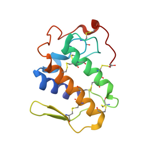
Structure of myotoxin II, a catalytically inactive Lys49 phospholipase A2 homologue from Atropoides nummifer venom.

Murakami, M.T.Melo, C.C.Angulo, Y.Lomonte, B.Arni, R.K.

(2006) Acta Crystallogr Sect F Struct Biol Cryst Commun 62: 423-426

  • DOI: https://doi.org/10.1107/S1744309106010700
  • Primary Citation of Related Structures:  
    2AOZ

  • PubMed Abstract: 

    Lys49 snake-venom phospholipase A2 (PLA2) homologues are highly myotoxic proteins which, although lacking catalytic activity, possess the ability to disrupt biological membranes, inducing significant muscle-tissue loss and permanent disability in severely envenomed patients. Since the structural basis for their toxic activity is still only partially understood, the structure of myotoxin II, a monomeric Lys49 PLA2 homologue from Atropoides nummifer, has been determined at 2.08 angstroms resolution and the anion-binding site has been characterized.


  • Organizational Affiliation

    Department of Physics, IBILCE/UNESP, São José do Rio Preto-SP, Brazil.


Macromolecules
Find similar proteins by:  (by identity cutoff)  |  3D Structure
Entity ID: 1
MoleculeChains Sequence LengthOrganismDetailsImage
Phospholipase A2 homolog121Atropoides nummiferMutation(s): 0 
UniProt
Find proteins for P82950 (Atropoides nummifer)
Explore P82950 
Go to UniProtKB:  P82950
Entity Groups  
Sequence Clusters30% Identity50% Identity70% Identity90% Identity95% Identity100% Identity
UniProt GroupP82950
Sequence Annotations
Expand
  • Reference Sequence
Small Molecules
Ligands 1 Unique
IDChains Name / Formula / InChI Key2D Diagram3D Interactions
SO4
Query on SO4

Download Ideal Coordinates CCD File 
B [auth A]SULFATE ION
O4 S
QAOWNCQODCNURD-UHFFFAOYSA-L
Experimental Data & Validation

Experimental Data

  • Method: X-RAY DIFFRACTION
  • Resolution: 2.08 Å
  • R-Value Free: 0.275 
  • R-Value Work: 0.219 
  • R-Value Observed: 0.221 
  • Space Group: P 43 21 2
Unit Cell:
Length ( Å )Angle ( ˚ )
a = 68.939α = 90
b = 68.939β = 90
c = 64.069γ = 90
Software Package:
Software NamePurpose
REFMACrefinement
DENZOdata reduction
SCALEPACKdata scaling
AMoREphasing

Structure Validation

View Full Validation Report


Entry History 

Deposition Data

Revision History  (Full details and data files)

  • Version 1.0: 2006-07-25
    Type: Initial release
  • Version 1.1: 2008-04-30
    Changes: Version format compliance
  • Version 1.2: 2011-07-13
    Changes: Advisory, Version format compliance
  • Version 1.3: 2023-08-23
    Changes: Data collection, Database references, Derived calculations, Refinement description