3M0I

Human Aldose Reductase mutant T113V in complex with Zopolrestat


Experimental Data Snapshot

  • Method: X-RAY DIFFRACTION
  • Resolution: 1.07 Å
  • R-Value Free: 0.145 
  • R-Value Observed: 0.115 

wwPDB Validation   3D Report Full Report


Ligand Structure Quality Assessment 


This is version 1.3 of the entry. See complete history


Literature

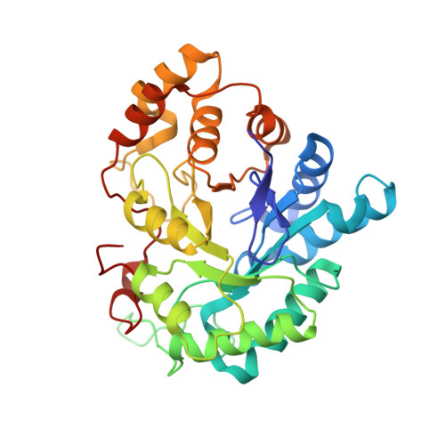
Ligand-induced fit affects binding modes and provokes changes in crystal packing of aldose reductase

Koch, C.Heine, A.Klebe, G.

(2011) Biochim Biophys Acta 1810: 879-887

  • DOI: https://doi.org/10.1016/j.bbagen.2011.06.001
  • Primary Citation of Related Structures:  
    3LEN, 3M0I, 3M64, 3MB9, 3MC5, 3P2V

  • PubMed Abstract: 

    Flexibility is a common feature of proteins. For human aldose reductase, a variety of conformers have been observed in crystalline complexes with different inhibitors. A study of crystal structures and isothermal titration calorimetry was performed on wild type and mutated aldose reductase. Though the interaction to the mutated residue Thr113 does not directly alter the binding mode of zopolrestat to aldose reductase, a shift of its basic scaffold is induced which affects the interaction with a flexible loop and introduces disorder. With the related inhibitor IDD393, two distinct binding site conformations result in two different crystal forms: While a backbone flip of the same residues as for zopolrestat is present in both crystal forms, a considerable side-chain movement of a phenylalanine is observed for only one crystal form. In consequence, residual mobility of adjacent amino acids is increased and some crystal contacts are prevented which reinforces different crystal packing. The structure of a benzothiazepine reveals a protein conformer, where this phenylalanine is further relocated resulting in the same altered crystal packing. Differences in the thermodynamic signature recorded for the various complexes relate to the structural differences. Crystal structures are accepted as "gold standard" for the interpretation of protein geometry, however, they are only one possible structure and can be influenced by crystal packing. In reverse, ligand binding can affect protein conformation and determine crystal packing. The phenomenon of such "polymorphic forms" is well appreciated, however rarely understood at the molecular level.


  • Organizational Affiliation

    Philipps-Universität, Department of Pharmaceutical Chemistry, Marburg, Germany.


Macromolecules
Find similar proteins by:  (by identity cutoff)  |  3D Structure
Entity ID: 1
MoleculeChains Sequence LengthOrganismDetailsImage
Aldose Reductase316Homo sapiensMutation(s): 1 
Gene Names: alr2
EC: 1.1.1.21
UniProt & NIH Common Fund Data Resources
Find proteins for P15121 (Homo sapiens)
Explore P15121 
Go to UniProtKB:  P15121
PHAROS:  P15121
GTEx:  ENSG00000085662 
Entity Groups  
Sequence Clusters30% Identity50% Identity70% Identity90% Identity95% Identity100% Identity
UniProt GroupP15121
Sequence Annotations
Expand
  • Reference Sequence
Small Molecules
Ligands 2 Unique
IDChains Name / Formula / InChI Key2D Diagram3D Interactions
NAP
Query on NAP

Download Ideal Coordinates CCD File 
B [auth A]NADP NICOTINAMIDE-ADENINE-DINUCLEOTIDE PHOSPHATE
C21 H28 N7 O17 P3
XJLXINKUBYWONI-NNYOXOHSSA-N
ZST
Query on ZST

Download Ideal Coordinates CCD File 
C [auth A]3,4-DIHYDRO-4-OXO-3-((5-TRIFLUOROMETHYL-2-BENZOTHIAZOLYL)METHYL)-1-PHTHALAZINE ACETIC ACID
C19 H12 F3 N3 O3 S
BCSVCWVQNOXFGL-UHFFFAOYSA-N
Binding Affinity Annotations 
IDSourceBinding Affinity
ZST BindingDB:  3M0I Ki: 19 (nM) from 1 assay(s)
IC50: min: 1.9, max: 81 (nM) from 17 assay(s)
Binding MOAD:  3M0I Kd: 1850 (nM) from 1 assay(s)
Experimental Data & Validation

Experimental Data

  • Method: X-RAY DIFFRACTION
  • Resolution: 1.07 Å
  • R-Value Free: 0.145 
  • R-Value Observed: 0.115 
  • Space Group: P 1
Unit Cell:
Length ( Å )Angle ( ˚ )
a = 40.04α = 76.19
b = 47.03β = 67.5
c = 47.4γ = 77.34
Software Package:
Software NamePurpose
MAR345dtbdata collection
CNSrefinement
SHELXL-97refinement
HKL-2000data reduction
HKL-2000data scaling
CNSphasing

Structure Validation

View Full Validation Report


Ligand Structure Quality Assessment 


Entry History 

Deposition Data

Revision History  (Full details and data files)

  • Version 1.0: 2011-03-09
    Type: Initial release
  • Version 1.1: 2011-07-13
    Changes: Version format compliance
  • Version 1.2: 2011-08-24
    Changes: Database references
  • Version 1.3: 2024-03-20
    Changes: Data collection, Database references, Derived calculations