3Q9A

Structure of neuronal nitric oxide synthase in the ferric state in complex with N-5-[2-(ethylsulfanyl)ethanimidoyl]-L-ornithine


Experimental Data Snapshot

  • Method: X-RAY DIFFRACTION
  • Resolution: 2.24 Å
  • R-Value Free: 0.236 
  • R-Value Work: 0.186 
  • R-Value Observed: 0.189 

wwPDB Validation   3D Report Full Report


Ligand Structure Quality Assessment 


This is version 1.3 of the entry. See complete history


Literature

Temperature-dependent spin crossover in neuronal nitric oxide synthase bound with the heme-coordinating thioether inhibitors.

Doukov, T.Li, H.Sharma, A.Martell, J.D.Soltis, S.M.Silverman, R.B.Poulos, T.L.

(2011) J Am Chem Soc 133: 8326-8334

  • DOI: https://doi.org/10.1021/ja201466v
  • Primary Citation of Related Structures:  
    3Q99, 3Q9A

  • PubMed Abstract: 

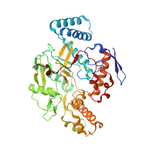
    A series of L-arginine analogue nitric oxide synthase inhibitors with a thioether tail have been shown to form an Fe-S thioether interaction as evidenced by continuous electron density between the Fe and S atoms. Even so, the Fe-S thioether interaction was found to be far less important for inhibitor binding than the hydrophobic interactions between the alkyl group in the thioether tail and surrounding protein (Martell et al. J. Am. Chem. Soc. 2010 , 132 , 798). However, among the few thioether inhibitors that showed Fe-S thioether interaction in crystal structures, variations in spin state (high-spin or low-spin) were observed dependent upon the heme iron oxidation state and temperature. Since modern synchrotron X-ray data collection is typically carried out at cryogenic temperatures, we reasoned that some of the discrepancies between cryo-crystal structures and room-temperature UV-visible spectroscopy could be the result of temperature-dependent spin-state changes. We, therefore, have characterized some of these neuronal nitric oxide synthase (nNOS)-thioether inhibitor complexes in both crystal and solution using EPR and UV-visible absorption spectrometry as a function of temperature and the heme iron redox state. We found that some thioether inhibitors switch from high to low spin at lower temperatures similar to the "spin crossover" phenomenon observed in many transition metal complexes.


  • Organizational Affiliation

    Macromolecular Crystallographic Group, Stanford Synchrotron Radiation Lightsource, SLAC, Stanford University, Stanford, California 94309, USA.


Macromolecules
Find similar proteins by:  (by identity cutoff)  |  3D Structure
Entity ID: 1
MoleculeChains Sequence LengthOrganismDetailsImage
Nitric oxide synthase, brain
A, B
422Rattus norvegicusMutation(s): 0 
Gene Names: Nos1Bnos
EC: 1.14.13.39
UniProt
Find proteins for P29476 (Rattus norvegicus)
Explore P29476 
Go to UniProtKB:  P29476
Entity Groups  
Sequence Clusters30% Identity50% Identity70% Identity90% Identity95% Identity100% Identity
UniProt GroupP29476
Sequence Annotations
Expand
  • Reference Sequence
Small Molecules
Ligands 5 Unique
IDChains Name / Formula / InChI Key2D Diagram3D Interactions
HEM
Query on HEM

Download Ideal Coordinates CCD File 
C [auth A],
H [auth B]
PROTOPORPHYRIN IX CONTAINING FE
C34 H32 Fe N4 O4
KABFMIBPWCXCRK-RGGAHWMASA-L
H4B
Query on H4B

Download Ideal Coordinates CCD File 
D [auth A],
I [auth B]
5,6,7,8-TETRAHYDROBIOPTERIN
C9 H15 N5 O3
FNKQXYHWGSIFBK-RPDRRWSUSA-N
JM7
Query on JM7

Download Ideal Coordinates CCD File 
F [auth A],
K [auth B]
N~5~-[2-(ethylsulfanyl)ethanimidoyl]-L-ornithine
C9 H19 N3 O2 S
GKZCWWBQARDOFD-ZETCQYMHSA-N
ZN
Query on ZN

Download Ideal Coordinates CCD File 
G [auth A]ZINC ION
Zn
PTFCDOFLOPIGGS-UHFFFAOYSA-N
ACT
Query on ACT

Download Ideal Coordinates CCD File 
E [auth A],
J [auth B]
ACETATE ION
C2 H3 O2
QTBSBXVTEAMEQO-UHFFFAOYSA-M
Experimental Data & Validation

Experimental Data

  • Method: X-RAY DIFFRACTION
  • Resolution: 2.24 Å
  • R-Value Free: 0.236 
  • R-Value Work: 0.186 
  • R-Value Observed: 0.189 
  • Space Group: P 21 21 21
Unit Cell:
Length ( Å )Angle ( ˚ )
a = 51.708α = 90
b = 110.607β = 90
c = 164.406γ = 90
Software Package:
Software NamePurpose
Blu-Icedata collection
REFMACrefinement
HKL-2000data reduction
SCALEPACKdata scaling
REFMACphasing

Structure Validation

View Full Validation Report


Ligand Structure Quality Assessment 


Entry History 

Deposition Data

Revision History  (Full details and data files)

  • Version 1.0: 2011-05-18
    Type: Initial release
  • Version 1.1: 2011-07-13
    Changes: Version format compliance
  • Version 1.2: 2017-11-08
    Changes: Refinement description
  • Version 1.3: 2023-09-13
    Changes: Data collection, Database references, Derived calculations, Refinement description