3R9B

Crystal structure of Mycobacterium smegmatis CYP164A2 in ligand free state


Experimental Data Snapshot

  • Method: X-RAY DIFFRACTION
  • Resolution: 1.89 Å
  • R-Value Free: 0.202 
  • R-Value Work: 0.183 
  • R-Value Observed: 0.184 

wwPDB Validation   3D Report Full Report


Ligand Structure Quality Assessment 


This is version 1.1 of the entry. See complete history


Literature

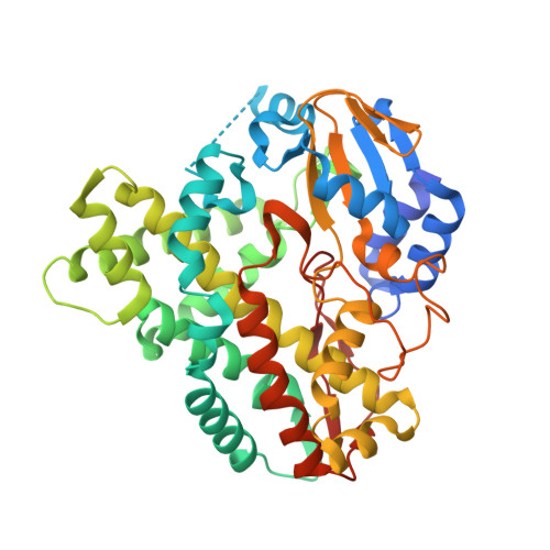
An enlarged, adaptable active site in CYP164 family P450 enzymes, the sole P450 in Mycobacterium leprae.

Agnew, C.R.Warrilow, A.G.Burton, N.M.Lamb, D.C.Kelly, S.L.Brady, R.L.

(2012) Antimicrob Agents Chemother 56: 391-402

  • DOI: https://doi.org/10.1128/AAC.05227-11
  • Primary Citation of Related Structures:  
    3R9B, 3R9C

  • PubMed Abstract: 

    CYP164 family P450 enzymes are found in only a subset of mycobacteria and include CYP164A1, which is the sole P450 found in Mycobacterium leprae, the causative agent of leprosy. This has previously led to interest in this enzyme as a potential drug target. Here we describe the first crystal structure of a CYP164 enzyme, CYP164A2 from Mycobacterium smegmatis. CYP164A2 has a distinctive, enlarged hydrophobic active site that extends above the porphyrin ring toward the access channels. Unusually, we find that CYP164A2 can simultaneously bind two econazole molecules in different regions of the enlarged active site and is accompanied by the rearrangement and ordering of the BC loop. The primary location is through a classic interaction of the azole group with the porphyrin iron. The second econazole molecule is bound to a unique site and is linked to a tetracoordinated metal ion complexed to one of the heme carboxylates and to the side chains of His 105 and His 364. All of these features are preserved in the closely homologous M. leprae CYP164A1. The computational docking of azole compounds to a homology model of CYP164A1 suggests that these compounds will form effective inhibitors and is supported by the correlation of parallel docking with experimental binding studies of CYP164A2. The binding of econazole to CYP164A2 occurs primarily through the high-spin "open" conformation of the enzyme (K(d) [dissociation constant] of 0.1 μM), with binding to the low-spin "closed" form being significantly hindered (K(d) of 338 μM). These studies support previous suggestions that azole derivatives may provide an effective strategy to improve the treatment of leprosy.


  • Organizational Affiliation

    School of Biochemistry, University of Bristol, Bristol, United Kingdom.


Macromolecules
Find similar proteins by:  (by identity cutoff)  |  3D Structure
Entity ID: 1
MoleculeChains Sequence LengthOrganismDetailsImage
CYTOCHROME P450 164A2
A, B, C
418Mycolicibacterium smegmatis MC2 155Mutation(s): 0 
Gene Names: MSMEG_6312
EC: 1.14
UniProt
Find proteins for A0R5U2 (Mycolicibacterium smegmatis (strain ATCC 700084 / mc(2)155))
Explore A0R5U2 
Go to UniProtKB:  A0R5U2
Entity Groups  
Sequence Clusters30% Identity50% Identity70% Identity90% Identity95% Identity100% Identity
UniProt GroupA0R5U2
Sequence Annotations
Expand
  • Reference Sequence
Small Molecules
Ligands 6 Unique
IDChains Name / Formula / InChI Key2D Diagram3D Interactions
HEM
Query on HEM

Download Ideal Coordinates CCD File 
D [auth A],
M [auth B],
V [auth C]
PROTOPORPHYRIN IX CONTAINING FE
C34 H32 Fe N4 O4
KABFMIBPWCXCRK-RGGAHWMASA-L
D12
Query on D12

Download Ideal Coordinates CCD File 
HA [auth C],
L [auth A],
U [auth B]
DODECANE
C12 H26
SNRUBQQJIBEYMU-UHFFFAOYSA-N
PO4
Query on PO4

Download Ideal Coordinates CCD File 
E [auth A],
F [auth A],
N [auth B],
W [auth C],
X [auth C]
PHOSPHATE ION
O4 P
NBIIXXVUZAFLBC-UHFFFAOYSA-K
EDO
Query on EDO

Download Ideal Coordinates CCD File 
AA [auth C]
BA [auth C]
CA [auth C]
DA [auth C]
EA [auth C]
AA [auth C],
BA [auth C],
CA [auth C],
DA [auth C],
EA [auth C],
G [auth A],
H [auth A],
K [auth A],
O [auth B],
P [auth B],
Q [auth B],
R [auth B],
S [auth B],
Y [auth C],
Z [auth C]
1,2-ETHANEDIOL
C2 H6 O2
LYCAIKOWRPUZTN-UHFFFAOYSA-N
K
Query on K

Download Ideal Coordinates CCD File 
GA [auth C],
J [auth A],
T [auth B]
POTASSIUM ION
K
NPYPAHLBTDXSSS-UHFFFAOYSA-N
NA
Query on NA

Download Ideal Coordinates CCD File 
FA [auth C],
I [auth A]
SODIUM ION
Na
FKNQFGJONOIPTF-UHFFFAOYSA-N
Experimental Data & Validation

Experimental Data

  • Method: X-RAY DIFFRACTION
  • Resolution: 1.89 Å
  • R-Value Free: 0.202 
  • R-Value Work: 0.183 
  • R-Value Observed: 0.184 
  • Space Group: C 1 2 1
Unit Cell:
Length ( Å )Angle ( ˚ )
a = 178.396α = 90
b = 102.776β = 90.06
c = 92.412γ = 90
Software Package:
Software NamePurpose
GDAdata collection
SHELXSphasing
REFMACrefinement
HKL-2000data reduction
SCALAdata scaling

Structure Validation

View Full Validation Report


Ligand Structure Quality Assessment 


Entry History 

Deposition Data

Revision History  (Full details and data files)

  • Version 1.0: 2012-04-04
    Type: Initial release
  • Version 1.1: 2024-03-20
    Changes: Data collection, Database references, Derived calculations, Refinement description