4UE8

Complex of D. melanogaster eIF4E with the 4E binding protein Thor


Experimental Data Snapshot

  • Method: X-RAY DIFFRACTION
  • Resolution: 1.10 Å
  • R-Value Free: 0.161 
  • R-Value Work: 0.145 
  • R-Value Observed: 0.146 

wwPDB Validation   3D Report Full Report


Ligand Structure Quality Assessment 


This is version 1.3 of the entry. See complete history


Literature

Molecular Architecture of 4E-BP Translational Inhibitors Bound to Eif4E.

Peter, D.Igreja, C.Weber, R.Wohlbold, L.Weiler, C.Ebertsch, L.Weichenrieder, O.Izaurralde, E.

(2015) Mol Cell 57: 1074

  • DOI: https://doi.org/10.1016/j.molcel.2015.01.017
  • Primary Citation of Related Structures:  
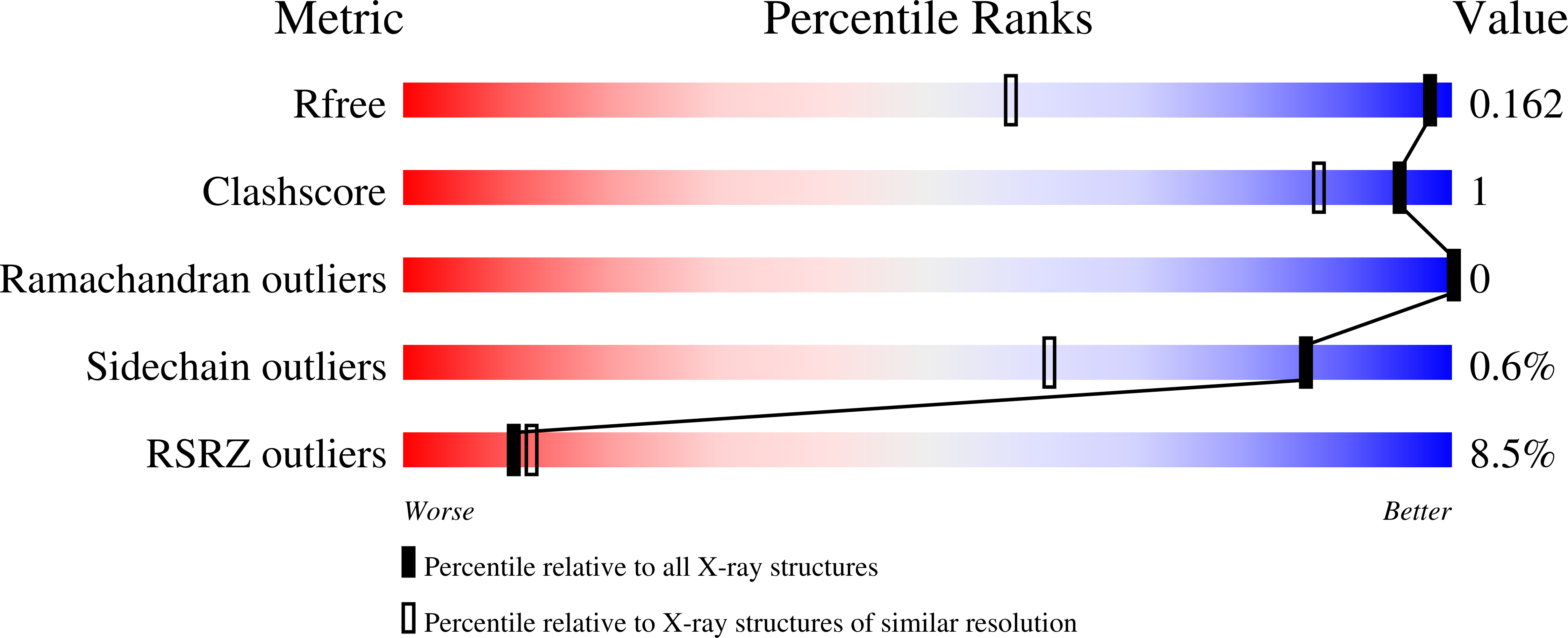
    4UE8, 4UE9, 4UEA, 4UEB, 4UEC, 4UED

  • PubMed Abstract: 

    The eIF4E-binding proteins (4E-BPs) represent a diverse class of translation inhibitors that are often deregulated in cancer cells. 4E-BPs inhibit translation by competing with eIF4G for binding to eIF4E through an interface that consists of canonical and non-canonical eIF4E-binding motifs connected by a linker. The lack of high-resolution structures including the linkers, which contain phosphorylation sites, limits our understanding of how phosphorylation inhibits complex formation. Furthermore, the binding mechanism of the non-canonical motifs is poorly understood. Here, we present structures of human eIF4E bound to 4E-BP1 and fly eIF4E bound to Thor, 4E-T, and eIF4G. These structures reveal architectural elements that are unique to 4E-BPs and provide insight into the consequences of phosphorylation. Guided by these structures, we designed and crystallized a 4E-BP mimic that shows increased repressive activity. Our studies pave the way for the rational design of 4E-BP mimics as therapeutic tools to decrease translation during oncogenic transformation.


  • Organizational Affiliation

    Department of Biochemistry, Max Planck Institute for Developmental Biology, Spemannstrasse 35, 72076 Tübingen, Germany.


Macromolecules
Find similar proteins by:  (by identity cutoff)  |  3D Structure
Entity ID: 1
MoleculeChains Sequence LengthOrganismDetailsImage
EUKARYOTIC TRANSLATION INITIATION FACTOR 4E184Drosophila melanogasterMutation(s): 0 
UniProt
Find proteins for P48598 (Drosophila melanogaster)
Explore P48598 
Go to UniProtKB:  P48598
Entity Groups  
Sequence Clusters30% Identity50% Identity70% Identity90% Identity95% Identity100% Identity
UniProt GroupP48598
Sequence Annotations
Expand
  • Reference Sequence
Find similar proteins by:  (by identity cutoff)  |  3D Structure
Entity ID: 2
MoleculeChains Sequence LengthOrganismDetailsImage
4E-BINDING PROTEIN THOR38Drosophila melanogasterMutation(s): 0 
UniProt
Find proteins for Q9XZ56 (Drosophila melanogaster)
Explore Q9XZ56 
Go to UniProtKB:  Q9XZ56
Entity Groups  
Sequence Clusters30% Identity50% Identity70% Identity90% Identity95% Identity100% Identity
UniProt GroupQ9XZ56
Sequence Annotations
Expand
  • Reference Sequence
Experimental Data & Validation

Experimental Data

  • Method: X-RAY DIFFRACTION
  • Resolution: 1.10 Å
  • R-Value Free: 0.161 
  • R-Value Work: 0.145 
  • R-Value Observed: 0.146 
  • Space Group: P 1 21 1
Unit Cell:
Length ( Å )Angle ( ˚ )
a = 35.45α = 90
b = 73.29β = 112.49
c = 38.26γ = 90
Software Package:
Software NamePurpose
PHENIXrefinement
XDSdata reduction
XSCALEdata scaling
PHASERphasing

Structure Validation

View Full Validation Report


Ligand Structure Quality Assessment 


Entry History 

Deposition Data

Revision History  (Full details and data files)

  • Version 1.0: 2015-02-25
    Type: Initial release
  • Version 1.1: 2015-03-04
    Changes: Database references
  • Version 1.2: 2015-04-15
    Changes: Database references
  • Version 1.3: 2023-12-20
    Changes: Data collection, Database references, Derived calculations, Other, Refinement description