4V33

Crystal structure of the putative polysaccharide deacetylase BA0330 from bacillus anthracis


Experimental Data Snapshot

  • Method: X-RAY DIFFRACTION
  • Resolution: 1.48 Å
  • R-Value Free: 0.175 
  • R-Value Work: 0.157 
  • R-Value Observed: 0.157 

wwPDB Validation   3D Report Full Report


This is version 1.3 of the entry. See complete history


Literature

Two Putative Polysaccharide Deacetylases are Required for Osmotic Stability and Cell Shape Maintenance in Bacillus Anthracis.

Arnaouteli, S.Giastas, P.Andreou, A.Tzanodaskalaki, M.Aldridge, C.Tzartos, S.J.Vollmer, W.Eliopoulos, E.Bouriotis, V.

(2015) J Biol Chem 290: 13465

  • DOI: https://doi.org/10.1074/jbc.M115.640029
  • Primary Citation of Related Structures:  
    4V33

  • PubMed Abstract: 

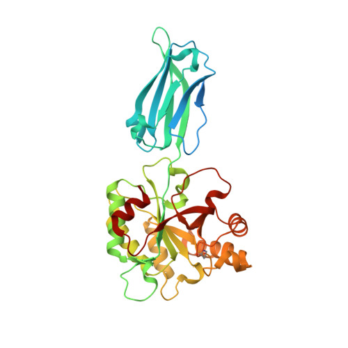
    Membrane-anchored lipoproteins have a broad range of functions and play key roles in several cellular processes in Gram-positive bacteria. BA0330 and BA0331 are the only lipoproteins among the 11 known or putative polysaccharide deacetylases of Bacillus anthracis. We found that both lipoproteins exhibit unique characteristics. BA0330 and BA0331 interact with peptidoglycan, and BA0330 is important for the adaptation of the bacterium to grow in the presence of a high concentration of salt, whereas BA0331 contributes to the maintenance of a uniform cell shape. They appear not to alter the peptidoglycan structure and do not contribute to lysozyme resistance. The high resolution x-ray structure of BA0330 revealed a C-terminal domain with the typical fold of a carbohydrate esterase 4 and an N-terminal domain unique for this family, composed of a two-layered (4 + 3) β-sandwich with structural similarity to fibronectin type 3 domains. Our data suggest that BA0330 and BA0331 have a structural role in stabilizing the cell wall of B. anthracis.


  • Organizational Affiliation

    From the Department of Biology, Enzyme Biotechnology Group, University of Crete, Vasilika Vouton, 70013 Heraklion, Crete, Greece.


Macromolecules
Find similar proteins by:  (by identity cutoff)  |  3D Structure
Entity ID: 1
MoleculeChains Sequence LengthOrganismDetailsImage
POLYSACCHARIDE DEACETYLASE-LIKE PROTEIN
A, B
360Bacillus anthracisMutation(s): 0 
UniProt
Find proteins for A0A6L8P8I1 (Bacillus anthracis)
Explore A0A6L8P8I1 
Go to UniProtKB:  A0A6L8P8I1
Entity Groups  
Sequence Clusters30% Identity50% Identity70% Identity90% Identity95% Identity100% Identity
UniProt GroupA0A6L8P8I1
Sequence Annotations
Expand
  • Reference Sequence
Small Molecules
Ligands 3 Unique
IDChains Name / Formula / InChI Key2D Diagram3D Interactions
ZN
Query on ZN

Download Ideal Coordinates CCD File 
D [auth A],
I [auth B]
ZINC ION
Zn
PTFCDOFLOPIGGS-UHFFFAOYSA-N
EDO
Query on EDO

Download Ideal Coordinates CCD File 
F [auth A],
G [auth A],
K [auth B],
L [auth B]
1,2-ETHANEDIOL
C2 H6 O2
LYCAIKOWRPUZTN-UHFFFAOYSA-N
ACT
Query on ACT

Download Ideal Coordinates CCD File 
C [auth A],
E [auth A],
H [auth B],
J [auth B]
ACETATE ION
C2 H3 O2
QTBSBXVTEAMEQO-UHFFFAOYSA-M
Modified Residues  1 Unique
IDChains TypeFormula2D DiagramParent
PXU
Query on PXU
A, B
L-PEPTIDE LINKINGC5 H9 N O3PRO
Experimental Data & Validation

Experimental Data

  • Method: X-RAY DIFFRACTION
  • Resolution: 1.48 Å
  • R-Value Free: 0.175 
  • R-Value Work: 0.157 
  • R-Value Observed: 0.157 
  • Space Group: C 1 2 1
Unit Cell:
Length ( Å )Angle ( ˚ )
a = 108.964α = 90
b = 68.763β = 96.86
c = 132.022γ = 90
Software Package:
Software NamePurpose
PHENIXrefinement
XDSdata reduction
SCALAdata scaling
PHASERphasing

Structure Validation

View Full Validation Report


Entry History 

Deposition Data

Revision History  (Full details and data files)

  • Version 1.0: 2015-04-08
    Type: Initial release
  • Version 1.1: 2015-04-15
    Changes: Database references
  • Version 1.2: 2015-06-10
    Changes: Database references
  • Version 1.3: 2024-01-10
    Changes: Data collection, Database references, Derived calculations, Other, Refinement description