


Crystal structure of human Thyroxine-binding globulin complexed with thyroine at 1.55 Angstrom resolution
Qi, X., Zhou, A.To be published.
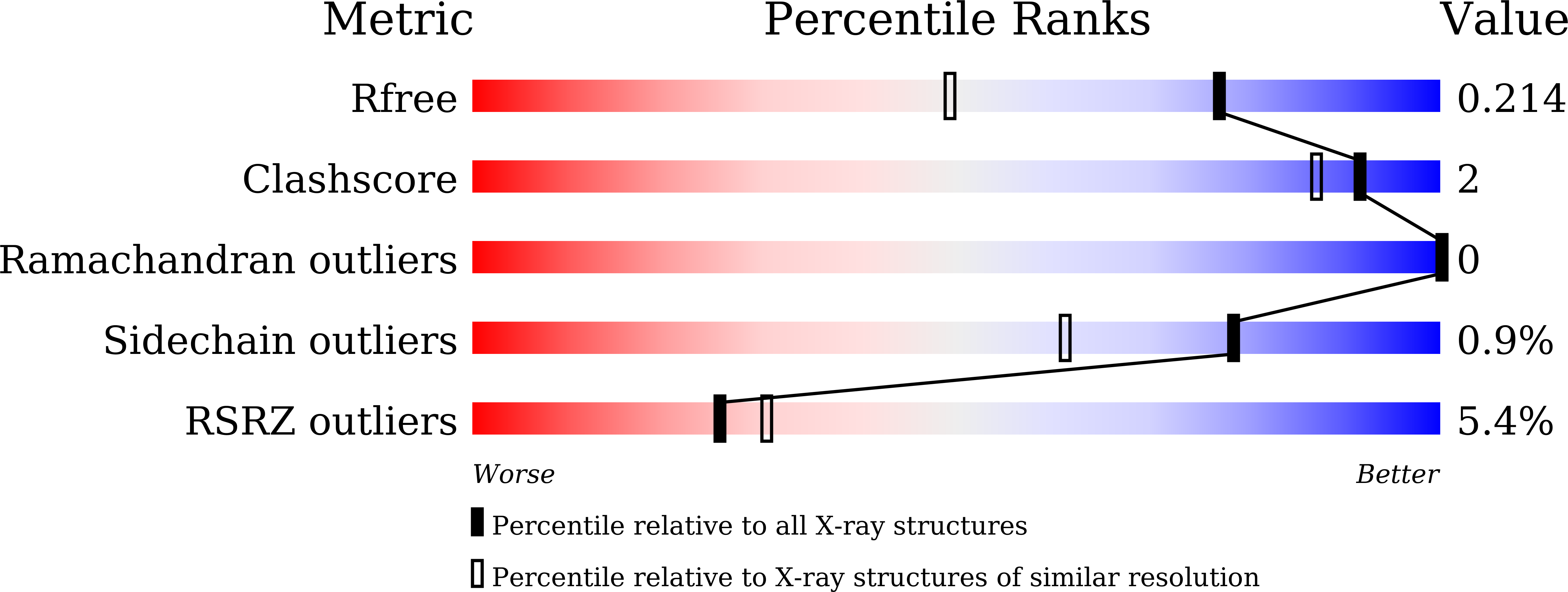
Experimental Data Snapshot
Entity ID: 1 | |||||
|---|---|---|---|---|---|
| Molecule | Chains | Sequence Length | Organism | Details | Image |
| Thyroxine-binding globulin | 395 | Homo sapiens | Mutation(s): 0 Gene Names: SERPINA7, TBG |  | |
UniProt & NIH Common Fund Data Resources | |||||
Find proteins for P05543 (Homo sapiens) Explore P05543 Go to UniProtKB: P05543 | |||||
PHAROS: P05543 GTEx: ENSG00000123561 | |||||
Entity Groups | |||||
| Sequence Clusters | 30% Identity50% Identity70% Identity90% Identity95% Identity100% Identity | ||||
| UniProt Group | P05543 | ||||
Sequence AnnotationsExpand | |||||
| |||||
| Ligands 5 Unique | |||||
|---|---|---|---|---|---|
| ID | Chains | Name / Formula / InChI Key | 2D Diagram | 3D Interactions | |
| T44 Query on T44 | B [auth A] | 3,5,3',5'-TETRAIODO-L-THYRONINE C15 H11 I4 N O4 XUIIKFGFIJCVMT-LBPRGKRZSA-N |  | ||
| GOL Query on GOL | C [auth A], D [auth A] | GLYCEROL C3 H8 O3 PEDCQBHIVMGVHV-UHFFFAOYSA-N |  | ||
| EDO Query on EDO | F [auth A] | 1,2-ETHANEDIOL C2 H6 O2 LYCAIKOWRPUZTN-UHFFFAOYSA-N |  | ||
| NA Query on NA | G [auth A] | SODIUM ION Na FKNQFGJONOIPTF-UHFFFAOYSA-N |  | ||
| F Query on F | E [auth A] | FLUORIDE ION F KRHYYFGTRYWZRS-UHFFFAOYSA-M |  | ||
| Length ( Å ) | Angle ( ˚ ) |
|---|---|
| a = 65.46 | α = 90 |
| b = 65.46 | β = 90 |
| c = 202.71 | γ = 90 |
| Software Name | Purpose |
|---|---|
| REFMAC | refinement |
| Coot | model building |
| MOSFLM | data processing |
| PHASER | phasing |
| SCALA | data reduction |
| SCALEPACK | data scaling |
| Funding Organization | Location | Grant Number |
|---|---|---|
| National Natural Science Foundation of China | China | 31170724 |
| nsfc | China | 31370727 |
RCSB PDB (citation) is hosted by
RCSB PDB is a member of the

