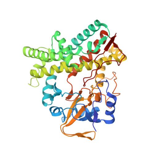
5UHU

Solution conformation of cytochrome P450 MycG with mycinamicin IV bound


Experimental Data Snapshot

  • Method: SOLUTION NMR
  • Conformers Calculated: 1200 
  • Conformers Submitted: 
  • Selection Criteria: structures with the least restraint violations 

wwPDB Validation   3D Report Full Report


This is version 1.3 of the entry. See complete history


Literature

Solution Conformations and Dynamics of Substrate-Bound Cytochrome P450 MycG.

Tietz, D.R.Podust, L.M.Sherman, D.H.Pochapsky, T.C.

(2017) Biochemistry 56: 2701-2714

  • DOI: https://doi.org/10.1021/acs.biochem.7b00291
  • Primary Citation of Related Structures:  
    5UHU

  • PubMed Abstract: 

    MycG is a P450 monooxygenase that catalyzes the sequential hydroxylation and epoxidation of mycinamicin IV (M-IV), the last two steps in the biosynthesis of mycinamicin II, a macrolide antibiotic isolated from Micromonospora griseorubida. The crystal structure of MycG with M-IV bound was previously determined but showed the bound substrate in an orientation that did not rationalize the observed regiochemistry of M-IV hydroxylation. Nuclear magnetic resonance paramagnetic relaxation enhancements provided evidence of an orientation of M-IV in the MycG active site more compatible with the observed chemistry, but substrate-induced changes in the enzyme structure were not characterized. We now describe the use of amide 1 H- 15 N residual dipolar couplings as experimental restraints in solvated "soft annealing" molecular dynamics simulations to generate solution structural ensembles of M-IV-bound MycG. Chemical shift perturbations, hydrogen-deuterium exchange, and 15 N relaxation behavior provide insight into the dynamic and electronic perturbations in the MycG structure in response to M-IV binding. The solution and crystallographic structures are compared, and the possibility that the crystallographic orientation of bound M-IV represents an inhibitory mode is discussed.


  • Organizational Affiliation

    Skaggs School of Pharmacy and Pharmaceutical Sciences, University of California , San Diego, California 92093, United States.


Macromolecules
Find similar proteins by:  (by identity cutoff)  |  3D Structure
Entity ID: 1
MoleculeChains Sequence LengthOrganismDetailsImage
Mycinamicin IV hydroxylase/epoxidase397Micromonospora griseorubidaMutation(s): 0 
Gene Names: mycG
EC: 1.14
UniProt
Find proteins for Q59523 (Micromonospora griseorubida)
Explore Q59523 
Go to UniProtKB:  Q59523
Entity Groups  
Sequence Clusters30% Identity50% Identity70% Identity90% Identity95% Identity100% Identity
UniProt GroupQ59523
Sequence Annotations
Expand
  • Reference Sequence
Small Molecules
Ligands 2 Unique
IDChains Name / Formula / InChI Key2D Diagram3D Interactions
MIV
Query on MIV

Download Ideal Coordinates CCD File 
C [auth A]MYCINAMICIN IV
C37 H61 N O11
DBTIHDIIXPQOFR-JMHKOBKLSA-N
HEM
Query on HEM

Download Ideal Coordinates CCD File 
B [auth A]PROTOPORPHYRIN IX CONTAINING FE
C34 H32 Fe N4 O4
KABFMIBPWCXCRK-RGGAHWMASA-L
Experimental Data & Validation

Experimental Data

  • Method: SOLUTION NMR
  • Conformers Calculated: 1200 
  • Conformers Submitted: 
  • Selection Criteria: structures with the least restraint violations 

Structure Validation

View Full Validation Report


Entry History & Funding Information

Deposition Data


Funding OrganizationLocationGrant Number
National Institutes of Health/National Institute of General Medical Sciences (NIH/NIGMS)United StatesR01GM44191

Revision History  (Full details and data files)

  • Version 1.0: 2017-08-23
    Type: Initial release
  • Version 1.1: 2017-09-27
    Changes: Author supporting evidence
  • Version 1.2: 2020-01-01
    Changes: Author supporting evidence, Data collection
  • Version 1.3: 2024-05-01
    Changes: Data collection, Database references