


Crystal structure of lysozyme from Equus asinus
Jiang, L., Liu, J.To be published.
Experimental Data Snapshot
wwPDB Validation 3D Report Full Report

Entity ID: 1 | |||||
|---|---|---|---|---|---|
| Molecule | Chains | Sequence Length | Organism | Details | Image |
| Lysozyme C | 129 | Equus asinus | Mutation(s): 0 EC: 3.2.1.17 | 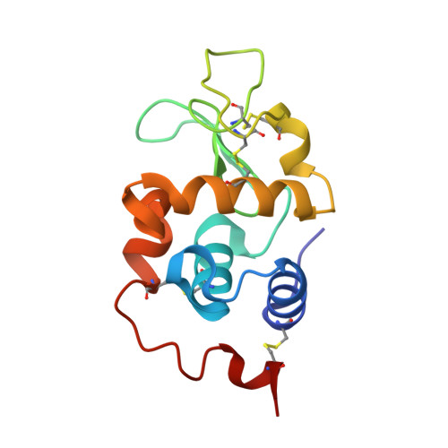 | |
UniProt | |||||
Find proteins for P11375 (Equus asinus) Explore P11375 Go to UniProtKB: P11375 | |||||
Entity Groups | |||||
| Sequence Clusters | 30% Identity50% Identity70% Identity90% Identity95% Identity100% Identity | ||||
| UniProt Group | P11375 | ||||
Sequence AnnotationsExpand | |||||
| |||||
| Ligands 1 Unique | |||||
|---|---|---|---|---|---|
| ID | Chains | Name / Formula / InChI Key | 2D Diagram | 3D Interactions | |
| CA (Subject of Investigation/LOI) Query on CA | C [auth A], D [auth B] | CALCIUM ION Ca BHPQYMZQTOCNFJ-UHFFFAOYSA-N |  | ||
| Length ( Å ) | Angle ( ˚ ) |
|---|---|
| a = 57.543 | α = 90 |
| b = 57.543 | β = 90 |
| c = 73.359 | γ = 120 |
| Software Name | Purpose |
|---|---|
| REFMAC | refinement |
| HKL-2000 | data reduction |
| HKL-2000 | data scaling |
| BALBES | phasing |
RCSB PDB (citation) is hosted by
RCSB PDB is a member of the

