Crystal structure of carboxylesterase from Pseudomonas fluorescens, an alpha/beta hydrolase with broad substrate specificity.
Kim, K.K., Song, H.K., Shin, D.H., Hwang, K.Y., Choe, S., Yoo, O.J., Suh, S.W.(1997) Structure 5: 1571-1584
- PubMed: 9438866
- DOI: https://doi.org/10.1016/s0969-2126(97)00306-7
- Primary Citation of Related Structures:
1AUO, 1AUR - PubMed Abstract:
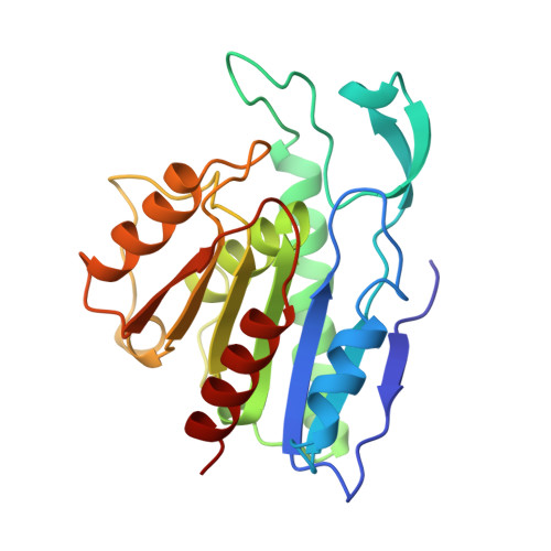
A group of esterases, classified as carboxylesterases, hydrolyze carboxylic ester bonds with relatively broad substrate specificity and are useful for stereospecific synthesis and hydrolysis of esters. One such carboxylesterase from Pseudomonas fluorescens is a homodimeric enzyme, consisting of 218-residue subunits. It shows a limited sequence similarity to some members of the alpha/beta hydrolase superfamily. Although crystal structures of a number of serine esterases and lipases have been reported, structural information on carboxylesterases is very limited. This study was undertaken in order to provide such information and to understand a structural basis for the substrate specificity of this carboxylesterase. In this study, the crystal structure of carboxylesterase from P. fluorescens has been determined by the isomorphous replacement method and refined to 1.8 A resolution. Each subunit consists of a central seven-stranded beta sheet flanked by six alpha helices. The structure reveals the catalytic triad as Ser 114-His 199-Asp 168. The structure of the enzyme in complex with the inhibitor phenylmethylsulfonyl fluoride has also been determined and refined to 2.5 . The inhibitor is covalently attached to Ser 114 of both subunits, with the aromatic ring occupying a hydrophobic site defined by the aliphatic sidechains of Leu23, Ile58, Ile70, Met73 and Val170. No large structural changes are observed between the free and inhibitor-bound structures. Carboxylesterase from P. fluorescens has the alpha/beta hydrolase fold and the Ser-His-Asp catalytic triad. The active-site cleft in each subunit is formed by the six loops covering the catalytic serine residue. Three of the active-site loops in each subunit are involved in a head-to-head subunit interaction to form a dimer; it may be these extra structural elements, not seen in other esterases, that account for the inability of carboxylesterase to hydrolyze long chain fatty acids. As a result of dimerization, the active-site clefts from the two subunits merge to form holes in the dimer. The active-site clefts are relatively open and thus the catalytic residues are exposed to the solvent. An oxyanion hole, formed by nitrogen atoms of Leu23 and Gln115, is present in both the free and inhibitor-bound structures. An open active site, as well as a large binding pocket for the acid part of substrates, in P. fluorescens carboxylesterase may contribute to its relatively broad substrate specificity.
Organizational Affiliation:
Department of Chemistry, College of Natural Sciences, Seoul National University, Korea.