2KT4

Lipocalin Q83 is a Siderocalin


Experimental Data Snapshot

  • Method: SOLUTION NMR
  • Conformers Calculated: 50 
  • Conformers Submitted: 20 
  • Selection Criteria: structures with the least restraint violations 

wwPDB Validation   3D Report Full Report


This is version 1.2 of the entry. See complete history


Literature

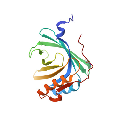
The v-myc-induced Q83 lipocalin is a siderocalin.

Coudevylle, N.Geist, L.Hotzinger, M.Hartl, M.Kontaxis, G.Bister, K.Konrat, R.

(2010) J Biol Chem 285: 41646-41652

  • DOI: https://doi.org/10.1074/jbc.M110.123331
  • Primary Citation of Related Structures:  
    2KT4

  • PubMed Abstract: 

    Siderocalins are atypical lipocalins able to capture siderophores with high affinity. They contribute to the innate immune response by interfering with bacterial siderophore-mediated iron uptake but are also involved in numerous physiological processes such as inflammation, iron delivery, tissue differentiation, and cancer progression. The Q83 lipocalin was originally identified based on its overexpression in quail embryo fibroblasts transformed by the v-myc oncogene. We show here that Q83 is a siderocalin, binding the siderophore enterobactin with an affinity and mode of binding nearly identical to that of neutrophil gelatinase-associated lipocalin (NGAL), the prototypical siderocalin. This strengthens the role of siderocalins in cancer progression and inflammation. In addition, we also present the solution structure of Q83 in complex with intact enterobactin and a detailed analysis of the Q83 binding mode, including mutagenesis of the critical residues involved in enterobactin binding. These data provide a first insight into the molecular details of siderophore binding and delineate the common molecular properties defining the siderocalin protein family.


  • Organizational Affiliation

    Department of Structural and Computational Biology, Max F Perutz Laboratories, University of Vienna, 1030 Vienna, Austria. nicolas.coudevylle@univie.ac.at


Macromolecules
Find similar proteins by:  (by identity cutoff)  |  3D Structure
Entity ID: 1
MoleculeChains Sequence LengthOrganismDetailsImage
Extracellular fatty acid-binding proteinA [auth B]157Coturnix japonicaMutation(s): 0 
Membrane Entity: Yes 
UniProt
Find proteins for Q9I9P7 (Coturnix japonica)
Explore Q9I9P7 
Go to UniProtKB:  Q9I9P7
Entity Groups  
Sequence Clusters30% Identity50% Identity70% Identity90% Identity95% Identity100% Identity
UniProt GroupQ9I9P7
Sequence Annotations
Expand
  • Reference Sequence
Small Molecules
Ligands 2 Unique
IDChains Name / Formula / InChI Key2D Diagram3D Interactions
EB4
Query on EB4

Download Ideal Coordinates CCD File 
B
N,N',N''-[(3S,7S,11S)-2,6,10-trioxo-1,5,9-trioxacyclododecane-3,7,11-triyl]tris(2,3-dihydroxybenzamide)
C30 H27 N3 O15
SERBHKJMVBATSJ-BZSNNMDCSA-N
GA
Query on GA

Download Ideal Coordinates CCD File 
C [auth B]GALLIUM (III) ION
Ga
CKHJYUSOUQDYEN-UHFFFAOYSA-N
Experimental Data & Validation

Experimental Data

  • Method: SOLUTION NMR
  • Conformers Calculated: 50 
  • Conformers Submitted: 20 
  • Selection Criteria: structures with the least restraint violations 

Structure Validation

View Full Validation Report


Entry History 

Deposition Data

Revision History  (Full details and data files)

  • Version 1.0: 2010-09-08
    Type: Initial release
  • Version 1.1: 2011-07-13
    Changes: Version format compliance
  • Version 1.2: 2016-01-27
    Changes: Database references