4GJJ

Crystal structure of Pseudomonas stutzeri L-rhamnose isomerase mutant H101N in complex with D-allopyranose


Experimental Data Snapshot

  • Method: X-RAY DIFFRACTION
  • Resolution: 2.38 Å
  • R-Value Free: 0.249 
  • R-Value Work: 0.196 
  • R-Value Observed: 0.196 

wwPDB Validation   3D Report Full Report


Ligand Structure Quality Assessment 


This is version 3.2 of the entry. See complete history


Literature

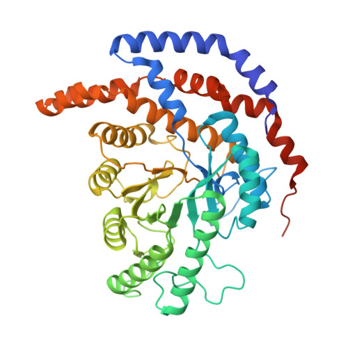
Structure of l-rhamnose isomerase in complex with l-rhamnopyranose demonstrates the sugar-ring opening mechanism and the role of a substrate sub-binding site.

Yoshida, H.Yoshihara, A.Teraoka, M.Yamashita, S.Izumori, K.Kamitori, S.

(2013) FEBS Open Bio 3: 35-40

  • DOI: https://doi.org/10.1016/j.fob.2012.11.008
  • Primary Citation of Related Structures:  
    4GJI, 4GJJ

  • PubMed Abstract: 

    l-Rhamnose isomerase (l-RhI) catalyzes the reversible isomerization of l-rhamnose to l-rhamnulose. Previously determined X-ray structures of l-RhI showed a hydride-shift mechanism for the isomerization of substrates in a linear form, but the mechanism for opening of the sugar-ring is still unclear. To elucidate this mechanism, we determined X-ray structures of a mutant l-RhI in complex with l-rhamnopyranose and d-allopyranose. Results suggest that a catalytic water molecule, which acts as an acid/base catalyst in the isomerization reaction, is likely to be involved in pyranose-ring opening, and that a newly found substrate sub-binding site in the vicinity of the catalytic site may recognize different anomers of substrates.


  • Organizational Affiliation

    Life Science Research Center and Faculty of Medicine, Kagawa University, 1750-1 Ikenobe, Miki-cho, Kita-gun, Kagawa 761-0793, Japan.


Macromolecules
Find similar proteins by:  (by identity cutoff)  |  3D Structure
Entity ID: 1
MoleculeChains Sequence LengthOrganismDetailsImage
L-rhamnose isomerase
A, B, C, D
438Stutzerimonas stutzeriMutation(s): 2 
Gene Names: L-RhI
EC: 5.3.1.14
UniProt
Find proteins for Q75WH8 (Stutzerimonas stutzeri)
Explore Q75WH8 
Go to UniProtKB:  Q75WH8
Entity Groups  
Sequence Clusters30% Identity50% Identity70% Identity90% Identity95% Identity100% Identity
UniProt GroupQ75WH8
Sequence Annotations
Expand
  • Reference Sequence
Small Molecules
Ligands 3 Unique
IDChains Name / Formula / InChI Key2D Diagram3D Interactions
AFD
Query on AFD

Download Ideal Coordinates CCD File 
K [auth B]alpha-D-allopyranose
C6 H12 O6
WQZGKKKJIJFFOK-UKFBFLRUSA-N
AOS
Query on AOS

Download Ideal Coordinates CCD File 
G [auth A],
N [auth C],
R [auth D]
D-ALLOSE
C6 H12 O6
GZCGUPFRVQAUEE-BGPJRJDNSA-N
MN
Query on MN

Download Ideal Coordinates CCD File 
E [auth A]
F [auth A]
H [auth B]
I [auth B]
J [auth B]
E [auth A],
F [auth A],
H [auth B],
I [auth B],
J [auth B],
L [auth C],
M [auth C],
O [auth D],
P [auth D],
Q [auth D]
MANGANESE (II) ION
Mn
WAEMQWOKJMHJLA-UHFFFAOYSA-N
Experimental Data & Validation

Experimental Data

  • Method: X-RAY DIFFRACTION
  • Resolution: 2.38 Å
  • R-Value Free: 0.249 
  • R-Value Work: 0.196 
  • R-Value Observed: 0.196 
  • Space Group: P 1 21 1
Unit Cell:
Length ( Å )Angle ( ˚ )
a = 74.65α = 90
b = 104.381β = 107.91
c = 113.972γ = 90
Software Package:
Software NamePurpose
ADSCdata collection
MOLREPphasing
CNSrefinement
HKL-2000data reduction
HKL-2000data scaling

Structure Validation

View Full Validation Report


Ligand Structure Quality Assessment 


Entry History 

Deposition Data

Revision History  (Full details and data files)

  • Version 1.0: 2012-12-12
    Type: Initial release
  • Version 1.1: 2014-05-28
    Changes: Database references
  • Version 2.0: 2017-11-15
    Changes: Advisory, Atomic model, Structure summary
  • Version 3.0: 2017-11-22
    Changes: Advisory, Atomic model, Derived calculations
  • Version 3.1: 2020-07-29
    Type: Remediation
    Reason: Carbohydrate remediation
    Changes: Data collection, Database references, Derived calculations, Structure summary
  • Version 3.2: 2023-11-08
    Changes: Data collection, Database references, Refinement description, Structure summary