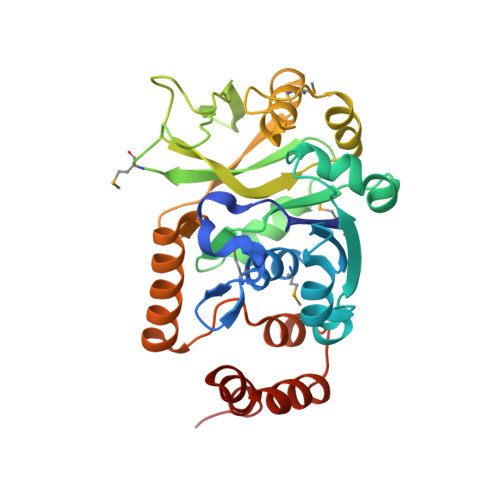
1MC3 | pdb_00001mc3

CRYSTAL STRUCTURE OF RFFH


Experimental Data Snapshot

  • Method: X-RAY DIFFRACTION
  • Resolution: 2.60 Å
  • R-Value Free: 
    0.280 (Depositor), 0.282 (DCC) 
  • R-Value Work: 
    0.223 (Depositor), 0.221 (DCC) 

wwPDB Validation   3D Report Full Report


Ligand Structure Quality Assessment 


This is version 1.5 of the entry. See complete history


Literature

Crystal Structure of Escherichia coli Glucose-1-Phosphate Thymidylyltransferase (RffH) Complexed with dTTP and Mg2+

Sivaraman, J.Sauve, V.Matte, A.Cygler, M.

(2002) J Biological Chem 277: 44214-44219

  • DOI: https://doi.org/10.1074/jbc.M206932200
  • Primary Citation of Related Structures:  
    1MC3

  • PubMed Abstract: 

    The enzyme glucose-1-phosphate thymidylyltransferase (RffH), the product of the rffh gene, catalyzes one of the steps in the synthesis of enterobacterial common antigen (ECA), a cell surface glycolipid found in Gram-negative enteric bacteria. In Escherichia coli two gene products, RffH and RmlA, catalyze the same enzymatic reaction and are homologous in sequence; however, they are part of different operons and function in different pathways. We report the crystal structure of RffH bound to deoxythymidine triphosphate (dTTP), the phosphate donor, and Mg(2+), refined at 2.6 A to an R-factor of 22.3% (R(free) = 28.4%). The crystal structure of RffH shows a tetrameric enzyme best described as a dimer of dimers. Each monomer has an overall alpha/beta fold and consists of two domains, a larger nucleotide binding domain (residues 1-115, 222-291) and a smaller sugar-binding domain (116-221), with the active site located at the domain interface. The Mg(2+) ion is coordinated by two conserved aspartates and the alpha-phosphate of deoxythymidine triphosphate. Its location corresponds well to that in a structurally similar domain of N-acetylglucosamine-1-phosphate uridylyltransferase (GlmU). Analysis of the RffH, RmlA, and GlmU complexes with substrates and products provides an explanation for their different affinities for Mg(2+) and leads to a proposal for the dynamics along the reaction pathway.


  • Organizational Affiliation

    Department of Biochemistry, McGill University, Montréal, Québec H3G 1Y6, Canada.


Macromolecules
Find similar proteins by:  (by identity cutoff)  |  3D Structure
Entity ID: 1
MoleculeChains Sequence LengthOrganismDetailsImage
GLUCOSE-1-PHOSPHATE THYMIDYLYLTRANSFERASE
A, B
296Escherichia coliMutation(s): 5 
EC: 2.7.7.24
UniProt
Find proteins for P61887 (Escherichia coli (strain K12))
Explore P61887 
Go to UniProtKB:  P61887
Entity Groups  
Sequence Clusters30% Identity50% Identity70% Identity90% Identity95% Identity100% Identity
UniProt GroupP61887
Sequence Annotations
Expand
  • Reference Sequence
Experimental Data & Validation

Experimental Data

  • Method: X-RAY DIFFRACTION
  • Resolution: 2.60 Å
  • R-Value Free:  0.280 (Depositor), 0.282 (DCC) 
  • R-Value Work:  0.223 (Depositor), 0.221 (DCC) 
Space Group: P 21 21 2
Unit Cell:
Length ( Å )Angle ( ˚ )
a = 144.338α = 90
b = 71.682β = 90
c = 59.413γ = 90
Software Package:
Software NamePurpose
SCALEPACKdata scaling
AMoREphasing
CNSrefinement

Structure Validation

View Full Validation Report


Ligand Structure Quality Assessment 


Entry History 

Deposition Data

Revision History  (Full details and data files)

  • Version 1.0: 2002-11-20
    Type: Initial release
  • Version 1.1: 2008-04-28
    Changes: Version format compliance
  • Version 1.2: 2011-07-13
    Changes: Version format compliance
  • Version 1.3: 2017-10-11
    Changes: Refinement description
  • Version 1.4: 2018-01-31
    Changes: Experimental preparation
  • Version 1.5: 2024-10-30
    Changes: Data collection, Database references, Derived calculations, Structure summary