1N83

Crystal Structure of the complex between the Orphan Nuclear Hormone Receptor ROR(alpha)-LBD and Cholesterol


Experimental Data Snapshot

  • Method: X-RAY DIFFRACTION
  • Resolution: 1.63 Å
  • R-Value Free: 0.223 
  • R-Value Work: 0.201 
  • R-Value Observed: 0.202 

wwPDB Validation   3D Report Full Report


Ligand Structure Quality Assessment 


This is version 1.3 of the entry. See complete history


Literature

X-ray Structure of hROR(alpha) LBD at 1.63A: Structural and Functional data that Cholesterol or a Cholesterol derivative is the natural ligand of ROR(alpha)

Kallen, J.A.Schlaeppi, J.M.Bitsch, F.Geisse, S.Geiser, M.Delhon, I.Fournier, B.

(2002) Structure 10: 1697-1702

  • DOI: https://doi.org/10.1016/s0969-2126(02)00912-7
  • Primary Citation of Related Structures:  
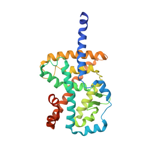
    1N83

  • PubMed Abstract: 

    The retinoic acid-related orphan receptor alpha (RORalpha) is an orphan member of the subfamily 1 of nuclear hormone receptors. No X-ray structure of RORalpha has been described so far, and no ligand has been identified. We describe the first crystal structure of the ligand binding domain (LBD) of RORalpha, at 1.63 A resolution. This structure revealed a ligand present in the ligand binding pocket (LBP), which was identified by X-ray crystallography as cholest-5-en-3beta-ol (cholesterol). Moreover, RORalpha transcriptional activity could be modulated by changes in intracellular cholesterol level or mutation of residues involved in cholesterol binding. These findings suggest that RORalpha could play a key role in the regulation of cholesterol homeostasis and thus represents an important drug target in cholesterol-related diseases.


  • Organizational Affiliation

    Central Technologies, Protein Structure Unit, Novartis Pharma AG, CH-4002 Basel, Switzerland. joerg.kallen@pharma.novartis.com


Macromolecules
Find similar proteins by:  (by identity cutoff)  |  3D Structure
Entity ID: 1
MoleculeChains Sequence LengthOrganismDetailsImage
Nuclear receptor ROR-alpha270Homo sapiensMutation(s): 0 
UniProt & NIH Common Fund Data Resources
Find proteins for P35398 (Homo sapiens)
Explore P35398 
Go to UniProtKB:  P35398
PHAROS:  P35398
GTEx:  ENSG00000069667 
Entity Groups  
Sequence Clusters30% Identity50% Identity70% Identity90% Identity95% Identity100% Identity
UniProt GroupP35398
Sequence Annotations
Expand
  • Reference Sequence
Small Molecules
Ligands 1 Unique
IDChains Name / Formula / InChI Key2D Diagram3D Interactions
CLR
Query on CLR

Download Ideal Coordinates CCD File 
B [auth A]CHOLESTEROL
C27 H46 O
HVYWMOMLDIMFJA-DPAQBDIFSA-N
Experimental Data & Validation

Experimental Data

  • Method: X-RAY DIFFRACTION
  • Resolution: 1.63 Å
  • R-Value Free: 0.223 
  • R-Value Work: 0.201 
  • R-Value Observed: 0.202 
  • Space Group: P 1 21 1
Unit Cell:
Length ( Å )Angle ( ˚ )
a = 54.8α = 90
b = 49.7β = 98.4
c = 60.5γ = 90
Software Package:
Software NamePurpose
DENZOdata reduction
SCALEPACKdata scaling
MLPHAREphasing
REFMACrefinement

Structure Validation

View Full Validation Report


Ligand Structure Quality Assessment 


Entry History 

Deposition Data

Revision History  (Full details and data files)

  • Version 1.0: 2002-12-11
    Type: Initial release
  • Version 1.1: 2008-04-28
    Changes: Version format compliance
  • Version 1.2: 2011-07-13
    Changes: Version format compliance
  • Version 1.3: 2024-02-14
    Changes: Data collection, Database references, Derived calculations